

homogenize the liquids and leave under the fume hood without shaking until the end of the 10 min (see Note 56).
3. Rinse cells with 0.01% Triton X-100 in 1Â PBS, gently manually shake the plate.
4. Wash with 0.01% Triton X-100 for 3 min. Repeat twice.
5. Incubate with 0.5% Triton X-100 for 10 min (see Note 57).
6. Incubate with 0.2 mg/mL RNase A solution for 30 min (see
Note 58).
7. Wash with PBS during 10 min. Repeat twice.
8. Incubate in 0.1 M HCl for 5 min (see Note 59).
9. Wash with 2Â SSC for 3 min. Repeat once.
10. Let stand in 50% formamide pH 7–7.2 at room temperature for
at least 1 h (see Notes 1 and 60).
3.3.3 Preparation
of the DNA-FISH Probes
1. In a microtube, mix 10–20 ng of labeled DNA-FISH probe (see
Note 61), 1 μL of SS-DNA from salmon testes and 6 μg or
12 μg (for BAC or fosmid probes, respectively) of human
Cot-1 DNA (see Note 62). Adjust the volume to 100 μL with
H 2 O.
2. Ethanol-precipitate the fluorescent probe mixture by adding
10 μL of 3 M sodium acetate pH 5.2 and 275 μL of cold
absolute ethanol.
3. Leave at À20
C for 1 h minimum (overnight is best). Centrifuge at 20,000 Â g for 30 min at 4
C.
4. Discard the supernatant (see Note 63), wash the pellet with
500 μL of 70% cold ethanol and centrifuge 15 min at
20,000 Â g at 4
C.
5. Discard the supernatant (see Note 63) and evaporate the residual liquid. The pellet must be dry.
6. Resuspend the nick-translated labeled DNA pellet in 5 μL
deionized formamide and let for 30 min at 37
C under an
aluminum foil while shaking on a thermomixer at 1000 rpm.
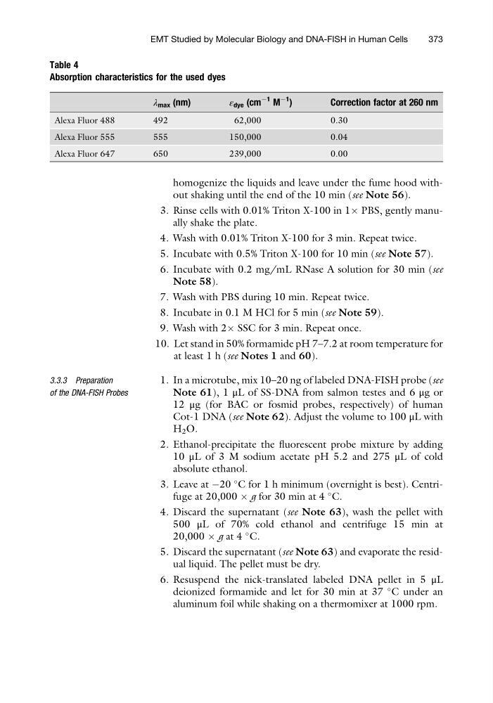
Table 4
Absorption characteristics for the used dyes
λ max (nm)
ε dye (cm
À1 M
À1
)
Correction factor at 260 nm
Alexa Fluor 488
492
62,000
0.30
Alexa Fluor 555
555
150,000
0.04
Alexa Fluor 647
650
239,000
0.00
EMT Studied by Molecular Biology and DNA-FISH in Human Cells
373
3. Rinse cells with 0.01% Triton X-100 in 1Â PBS, gently manually shake the plate.
4. Wash with 0.01% Triton X-100 for 3 min. Repeat twice.
5. Incubate with 0.5% Triton X-100 for 10 min (see Note 57).
6. Incubate with 0.2 mg/mL RNase A solution for 30 min (see
Note 58).
7. Wash with PBS during 10 min. Repeat twice.
8. Incubate in 0.1 M HCl for 5 min (see Note 59).
9. Wash with 2Â SSC for 3 min. Repeat once.
10. Let stand in 50% formamide pH 7–7.2 at room temperature for
at least 1 h (see Notes 1 and 60).
3.3.3 Preparation
of the DNA-FISH Probes
1. In a microtube, mix 10–20 ng of labeled DNA-FISH probe (see
Note 61), 1 μL of SS-DNA from salmon testes and 6 μg or
12 μg (for BAC or fosmid probes, respectively) of human
Cot-1 DNA (see Note 62). Adjust the volume to 100 μL with
H 2 O.
2. Ethanol-precipitate the fluorescent probe mixture by adding
10 μL of 3 M sodium acetate pH 5.2 and 275 μL of cold
absolute ethanol.
3. Leave at À20
C for 1 h minimum (overnight is best). Centrifuge at 20,000 Â g for 30 min at 4
C.
4. Discard the supernatant (see Note 63), wash the pellet with
500 μL of 70% cold ethanol and centrifuge 15 min at
20,000 Â g at 4
C.
5. Discard the supernatant (see Note 63) and evaporate the residual liquid. The pellet must be dry.
6. Resuspend the nick-translated labeled DNA pellet in 5 μL
deionized formamide and let for 30 min at 37
C under an
aluminum foil while shaking on a thermomixer at 1000 rpm.
Table 4
Absorption characteristics for the used dyes
λ max (nm)
ε dye (cm
À1 M
À1
)
Correction factor at 260 nm
Alexa Fluor 488
492
62,000
0.30
Alexa Fluor 555
555
150,000
0.04
Alexa Fluor 647
650
239,000
0.00
EMT Studied by Molecular Biology and DNA-FISH in Human Cells
373