

3.1.4 Harvesting Cells
for RT-qPCR
1. Heat 1Â DPBS and MCF10A growing media at 37
C and
Trypsin at room temperature.
2. Rinse cells with 10 mL of 1Â DPBS.
3. Remove the DPBS and add 1 mL of Trypsin for a 10 mm cell
culture dish and incubate 20–25 min at 37
C.
4. Gently hit the border of the cell culture dish to detach cells.
5. Add 9 mL of growing media and pipette up and down to
detach the cells sticking to the container.
6. Count the cells (see Note 23).
7. Transfer the 10 mL cell solution in a 50 mL tube and centrifuge
at 300 Â g for 5 min.
8. Discard the supernatant, and resuspend cell pellet in a volume
of 1Â DPBS in order to reach a cell concentration of 1.5 Â 10
6
cells/mL.
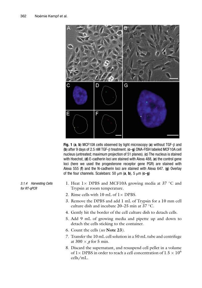
Fig. 1 (a, b) MCF10A cells observed by light microscopy (a) without TGF-β and
(b) after 9 days of 2.5 nM TGF-β treatment. (c–g) DNA-FISH labeled MCF10A cell
nucleus (untreated; maximum projection of 51 planes). (c) The nucleus is stained
with Hoechst, (d) E-cadherin loci are stained with Alexa 488, (e) the control gene
loci (here we used the progesterone receptor gene PGR) are stained with
Alexa 555 (f) and the N-cadherin loci are stained with Alexa 647. (g) Overlay
of the four channels. Scalebars: 50 μm (a, b), 5 μm (c–g)
362
Noe ´ mie Kempf et al.
for RT-qPCR
1. Heat 1Â DPBS and MCF10A growing media at 37
C and
Trypsin at room temperature.
2. Rinse cells with 10 mL of 1Â DPBS.
3. Remove the DPBS and add 1 mL of Trypsin for a 10 mm cell
culture dish and incubate 20–25 min at 37
C.
4. Gently hit the border of the cell culture dish to detach cells.
5. Add 9 mL of growing media and pipette up and down to
detach the cells sticking to the container.
6. Count the cells (see Note 23).
7. Transfer the 10 mL cell solution in a 50 mL tube and centrifuge
at 300 Â g for 5 min.
8. Discard the supernatant, and resuspend cell pellet in a volume
of 1Â DPBS in order to reach a cell concentration of 1.5 Â 10
6
cells/mL.
Fig. 1 (a, b) MCF10A cells observed by light microscopy (a) without TGF-β and
(b) after 9 days of 2.5 nM TGF-β treatment. (c–g) DNA-FISH labeled MCF10A cell
nucleus (untreated; maximum projection of 51 planes). (c) The nucleus is stained
with Hoechst, (d) E-cadherin loci are stained with Alexa 488, (e) the control gene
loci (here we used the progesterone receptor gene PGR) are stained with
Alexa 555 (f) and the N-cadherin loci are stained with Alexa 647. (g) Overlay
of the four channels. Scalebars: 50 μm (a, b), 5 μm (c–g)
362
Noe ´ mie Kempf et al.