

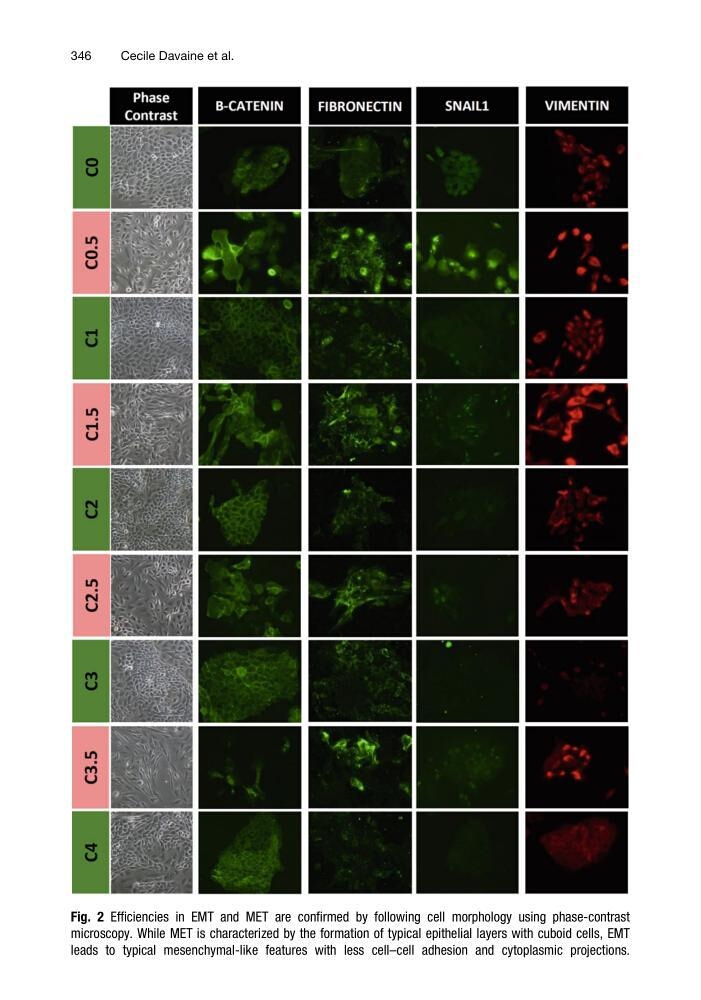
Fig. 2 Efficiencies in EMT and MET are confirmed by following cell morphology using phase-contrast
microscopy. While MET is characterized by the formation of typical epithelial layers with cuboid cells, EMT
leads to typical mesenchymal-like features with less cell–cell adhesion and cytoplasmic projections.
346
Cecile Davaine et al.
microscopy. While MET is characterized by the formation of typical epithelial layers with cuboid cells, EMT
leads to typical mesenchymal-like features with less cell–cell adhesion and cytoplasmic projections.
346
Cecile Davaine et al.