

transduction, ability to form tough and resistant layers) during
morphogenesis.
Similarly, succession of EM/ME transitions have also been
described during cancer progression [8, 9] and are crucial for
cancer cells dissemination and metastasis formation [10]. While
the tumoral microenvironment is highly dynamic and can favour
either EMT or MET, it is possible that pro-EMT and pro-MET
signals are successively received by cancer cells and can eventually
modify their biological properties. Indeed, activation of
EMT-transcription factors (EMT-TFs) controls a pleiotropic set
of cellular functions (cell proliferation, stemness, cell adhesion,
cytoskeleton dynamics, cell polarity, etc.) and can lead to a gradation of cellular states [1].
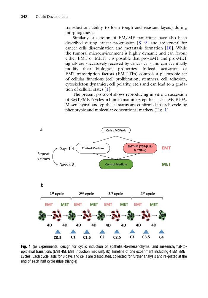
The present protocol allows reproducing in vitro a succession
of EMT/MET cycles in human mammary epithelial cells MCF10A.
Mesenchymal and epithelial status are confirmed in each cycle by
phenotypic and molecular conventional markers (Fig. 1).
Cells : MCF10A
Control Medium
EMT-IM (TGF-β, IL6, TNF-α)
Control Medium
EMT
MET
Days 1-4
Days 4-8
Repeat
x times
a
b
Fig. 1 (a) Experimental design for cyclic induction of epithelial-to-mesenchymal and mesenchymal-toepithelial transitions (EMT-IM: EMT induction medium). (b) Timeline of one experiment including 4 EMT/MET
cycles. Each cycle lasts for 8 days and cells are dissociated, collected for further analysis and re-plated at the
end of each half cycle (blue triangle)
342
Cecile Davaine et al.
morphogenesis.
Similarly, succession of EM/ME transitions have also been
described during cancer progression [8, 9] and are crucial for
cancer cells dissemination and metastasis formation [10]. While
the tumoral microenvironment is highly dynamic and can favour
either EMT or MET, it is possible that pro-EMT and pro-MET
signals are successively received by cancer cells and can eventually
modify their biological properties. Indeed, activation of
EMT-transcription factors (EMT-TFs) controls a pleiotropic set
of cellular functions (cell proliferation, stemness, cell adhesion,
cytoskeleton dynamics, cell polarity, etc.) and can lead to a gradation of cellular states [1].
The present protocol allows reproducing in vitro a succession
of EMT/MET cycles in human mammary epithelial cells MCF10A.
Mesenchymal and epithelial status are confirmed in each cycle by
phenotypic and molecular conventional markers (Fig. 1).
Cells : MCF10A
Control Medium
EMT-IM (TGF-β, IL6, TNF-α)
Control Medium
EMT
MET
Days 1-4
Days 4-8
Repeat
x times
a
b
Fig. 1 (a) Experimental design for cyclic induction of epithelial-to-mesenchymal and mesenchymal-toepithelial transitions (EMT-IM: EMT induction medium). (b) Timeline of one experiment including 4 EMT/MET
cycles. Each cycle lasts for 8 days and cells are dissociated, collected for further analysis and re-plated at the
end of each half cycle (blue triangle)
342
Cecile Davaine et al.