

continuous monolayers with large nuclei. Neural plate explants
remain as a tight cluster and project axons within few hours of
cell culture. Mesodermal and endodermal cells are significantly
larger and whiter than neural crest cells. They contain large
vitelline platelets.
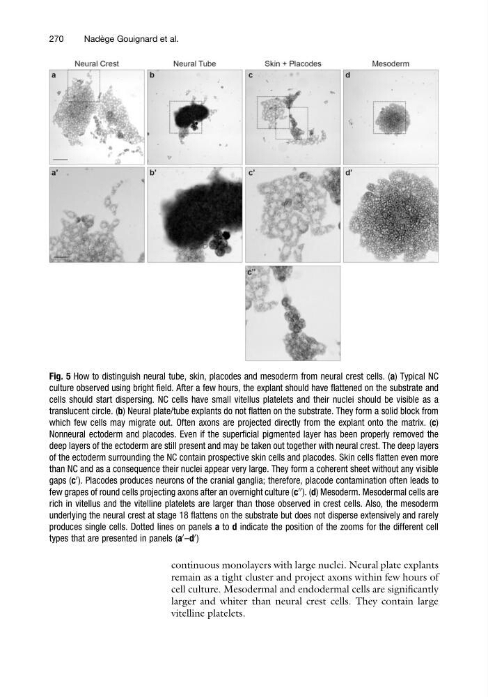
Fig. 5 How to distinguish neural tube, skin, placodes and mesoderm from neural crest cells. (a) Typical NC
culture observed using bright field. After a few hours, the explant should have flattened on the substrate and
cells should start dispersing. NC cells have small vitellus platelets and their nuclei should be visible as a
translucent circle. (b) Neural plate/tube explants do not flatten on the substrate. They form a solid block from
which few cells may migrate out. Often axons are projected directly from the explant onto the matrix. (c)
Nonneural ectoderm and placodes. Even if the superficial pigmented layer has been properly removed the
deep layers of the ectoderm are still present and may be taken out together with neural crest. The deep layers
of the ectoderm surrounding the NC contain prospective skin cells and placodes. Skin cells flatten even more
than NC and as a consequence their nuclei appear very large. They form a coherent sheet without any visible
gaps (c
0 ). Placodes produces neurons of the cranial ganglia; therefore, placode contamination often leads to
few grapes of round cells projecting axons after an overnight culture (c
00 ). (d) Mesoderm. Mesodermal cells are
rich in vitellus and the vitelline platelets are larger than those observed in crest cells. Also, the mesoderm
underlying the neural crest at stage 18 flattens on the substrate but does not disperse extensively and rarely
produces single cells. Dotted lines on panels a to d indicate the position of the zooms for the different cell
types that are presented in panels (a
0 –d
0 )
270
Nade ` ge Gouignard et al.
remain as a tight cluster and project axons within few hours of
cell culture. Mesodermal and endodermal cells are significantly
larger and whiter than neural crest cells. They contain large
vitelline platelets.
Fig. 5 How to distinguish neural tube, skin, placodes and mesoderm from neural crest cells. (a) Typical NC
culture observed using bright field. After a few hours, the explant should have flattened on the substrate and
cells should start dispersing. NC cells have small vitellus platelets and their nuclei should be visible as a
translucent circle. (b) Neural plate/tube explants do not flatten on the substrate. They form a solid block from
which few cells may migrate out. Often axons are projected directly from the explant onto the matrix. (c)
Nonneural ectoderm and placodes. Even if the superficial pigmented layer has been properly removed the
deep layers of the ectoderm are still present and may be taken out together with neural crest. The deep layers
of the ectoderm surrounding the NC contain prospective skin cells and placodes. Skin cells flatten even more
than NC and as a consequence their nuclei appear very large. They form a coherent sheet without any visible
gaps (c
0 ). Placodes produces neurons of the cranial ganglia; therefore, placode contamination often leads to
few grapes of round cells projecting axons after an overnight culture (c
00 ). (d) Mesoderm. Mesodermal cells are
rich in vitellus and the vitelline platelets are larger than those observed in crest cells. Also, the mesoderm
underlying the neural crest at stage 18 flattens on the substrate but does not disperse extensively and rarely
produces single cells. Dotted lines on panels a to d indicate the position of the zooms for the different cell
types that are presented in panels (a
0 –d
0 )
270
Nade ` ge Gouignard et al.