

calculate the appropriate volume, take into account that 250 μl
of cell-containing solutions are used for each microwell and
that cell numbers should be not less than 2 Â 10
5 cells and not
more than of 10
6 cells per microwell.
8. Stick the appropriate number of PDMS microwells (depending
on cell yields) to the bottom of a 6-well plate by using a
PBS drop.
9. Place 250 μl of cell solution on the microwells covering the
whole surface and centrifuge the plate for 2 min at 10 Â g.
10. Add 3 ml of organoid medium to the wells without disturbing
the PDMS surface.
Epithelial
Cells
ECM
proteins
Patterned
surface
A
XZ view
YZ view
DAPI/E-cadherin/K14
Top view
Middle view
Bottom view
DAPI/E-cadherin/K14
XZ view
YZ view
KEY
Matrigel
DAPI/E-cadherin/K14/F-actin
C
B
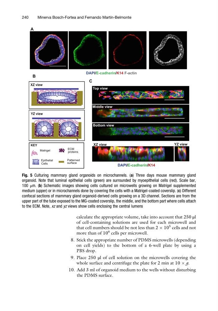
Fig. 5 Culturing mammary gland organoids on microchannels. (a) Three days mouse mammary gland
organoid. Note that luminal epithelial cells (green) are surrounded by myoepithelial cells (red). Scale bar,
100 μm. (b) Schematic images showing cells cultured on microwells growing on Matrigel supplemented
medium (upper) or in microchannels done by covering the cells with a Matrigel-coated coverslip. (c) Different
confocal sections of mammary gland organoid-derived cells growing on a 3D channel. Sections are from the
upper part of the tube exposed to the MG-coated coverslip, the middle, and the bottom part where cells attach
to the ECM. Note, xz and yz views show cells enclosing the central lumens
240
Minerva Bosch-Fortea and Fernando Martı ´n-Belmonte
of cell-containing solutions are used for each microwell and
that cell numbers should be not less than 2 Â 10
5 cells and not
more than of 10
6 cells per microwell.
8. Stick the appropriate number of PDMS microwells (depending
on cell yields) to the bottom of a 6-well plate by using a
PBS drop.
9. Place 250 μl of cell solution on the microwells covering the
whole surface and centrifuge the plate for 2 min at 10 Â g.
10. Add 3 ml of organoid medium to the wells without disturbing
the PDMS surface.
Epithelial
Cells
ECM
proteins
Patterned
surface
A
XZ view
YZ view
DAPI/E-cadherin/K14
Top view
Middle view
Bottom view
DAPI/E-cadherin/K14
XZ view
YZ view
KEY
Matrigel
DAPI/E-cadherin/K14/F-actin
C
B
Fig. 5 Culturing mammary gland organoids on microchannels. (a) Three days mouse mammary gland
organoid. Note that luminal epithelial cells (green) are surrounded by myoepithelial cells (red). Scale bar,
100 μm. (b) Schematic images showing cells cultured on microwells growing on Matrigel supplemented
medium (upper) or in microchannels done by covering the cells with a Matrigel-coated coverslip. (c) Different
confocal sections of mammary gland organoid-derived cells growing on a 3D channel. Sections are from the
upper part of the tube exposed to the MG-coated coverslip, the middle, and the bottom part where cells attach
to the ECM. Note, xz and yz views show cells enclosing the central lumens
240
Minerva Bosch-Fortea and Fernando Martı ´n-Belmonte