

vacuum using a vacuum desiccator for 20–30 min to degas the
mixture. The amount of PDMS should be determined depending on the number of substrates to fabricate.
10. Pour the PDMS over the silicon wafer or counter-mold and
bake at 80
C for 2 h in the oven.
11. Peel off the cured PDMS and cut it into small squared pieces
containing the patterned regions. Keep them on a 6-well plate
with the patterned region up-side.
12. Prepare 20 μg/ml Rhodamine-fibronectin solutions in PBS
using sterile conditions.
13. Coat the 3D microwells with the ECM solution by covering
the patterned region with a 300 μl drop. Incubate for 1 h at RT
in the dark.
14. Rinse the 3D microwells two times with sterile PBS and make
the last wash with milliQ water.
Epithelial
Cells
C
Top
Bottom
0
20
40
60
80
100
Proliferating cells (%)
**
KEY
Matrigel
ECM
proteins
Patterned
surface
2D micropatterns
3D micropatterns
A
0
20
40
Prolifer f f a
yz view
DAPI/Podocalyxin/Ki67
Top view
Middle view
Bottom view
B 1. Silicon wafer
2. PDMS mould
3. ECM Coating
4. Surface ECM removal
5. Cell seeding
7. Cell growth
6. Matrigel coating
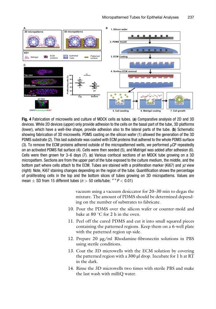
Fig. 4 Fabrication of microwells and culture of MDCK cells as tubes. (a) Comparative analysis of 2D and 3D
devices. While 2D devices (upper) only provide adhesion to the cells on the basal part of the tube, 3D platforms
(lower), which have a well-like shape, provide adhesion also to the lateral parts of the tube. (b) Schematic
showing fabrication of 3D microwells. PDMS casting on the silicon wafer (1) allowed the generation of the 3D
PDMS substrate (2). This last substrate was coated with ECM proteins that adhered to the whole PDMS surface
(3). To remove the ECM proteins adhered outside of the micropatterned wells, we performed μCP repeatedly
on an activated PDMS flat surface (4). Cells were then seeded (5), and Matrigel was added after adhesion (6).
Cells were then grown for 3–6 days (7). (c) Various confocal sections of an MDCK tube growing on a 3D
micropattern. Sections are from the upper part of the tube exposed to the culture medium, the middle, and the
bottom part where cells attach to the ECM. Tubes are stained with a proliferation marker (Ki67) and yz view
(right): Note, Ki67 staining changes depending on the region of the tube. Quantification shows the percentage
of proliferating cells in the top and the bottom slices of tubes growing on 3D micropatterns. Values are
mean Æ SD from 15 different tubes (n > 50 cells/tube;
∗∗
P < 0.01)
Micropatterned Tubes for Epithelial Analyses
237
mixture. The amount of PDMS should be determined depending on the number of substrates to fabricate.
10. Pour the PDMS over the silicon wafer or counter-mold and
bake at 80
C for 2 h in the oven.
11. Peel off the cured PDMS and cut it into small squared pieces
containing the patterned regions. Keep them on a 6-well plate
with the patterned region up-side.
12. Prepare 20 μg/ml Rhodamine-fibronectin solutions in PBS
using sterile conditions.
13. Coat the 3D microwells with the ECM solution by covering
the patterned region with a 300 μl drop. Incubate for 1 h at RT
in the dark.
14. Rinse the 3D microwells two times with sterile PBS and make
the last wash with milliQ water.
Epithelial
Cells
C
Top
Bottom
0
20
40
60
80
100
Proliferating cells (%)
**
KEY
Matrigel
ECM
proteins
Patterned
surface
2D micropatterns
3D micropatterns
A
0
20
40
Prolifer f f a
yz view
DAPI/Podocalyxin/Ki67
Top view
Middle view
Bottom view
B 1. Silicon wafer
2. PDMS mould
3. ECM Coating
4. Surface ECM removal
5. Cell seeding
7. Cell growth
6. Matrigel coating
Fig. 4 Fabrication of microwells and culture of MDCK cells as tubes. (a) Comparative analysis of 2D and 3D
devices. While 2D devices (upper) only provide adhesion to the cells on the basal part of the tube, 3D platforms
(lower), which have a well-like shape, provide adhesion also to the lateral parts of the tube. (b) Schematic
showing fabrication of 3D microwells. PDMS casting on the silicon wafer (1) allowed the generation of the 3D
PDMS substrate (2). This last substrate was coated with ECM proteins that adhered to the whole PDMS surface
(3). To remove the ECM proteins adhered outside of the micropatterned wells, we performed μCP repeatedly
on an activated PDMS flat surface (4). Cells were then seeded (5), and Matrigel was added after adhesion (6).
Cells were then grown for 3–6 days (7). (c) Various confocal sections of an MDCK tube growing on a 3D
micropattern. Sections are from the upper part of the tube exposed to the culture medium, the middle, and the
bottom part where cells attach to the ECM. Tubes are stained with a proliferation marker (Ki67) and yz view
(right): Note, Ki67 staining changes depending on the region of the tube. Quantification shows the percentage
of proliferating cells in the top and the bottom slices of tubes growing on 3D micropatterns. Values are
mean Æ SD from 15 different tubes (n > 50 cells/tube;
∗∗
P < 0.01)
Micropatterned Tubes for Epithelial Analyses
237