

5. Insect pins.
6. Fine incision scalpels (pfm Medical ophthalmic blades cat.
no. 200300715 or equivalent).
7. Low melting point agarose.
8. Micro-pipette with fine tips and 20 μL volume.
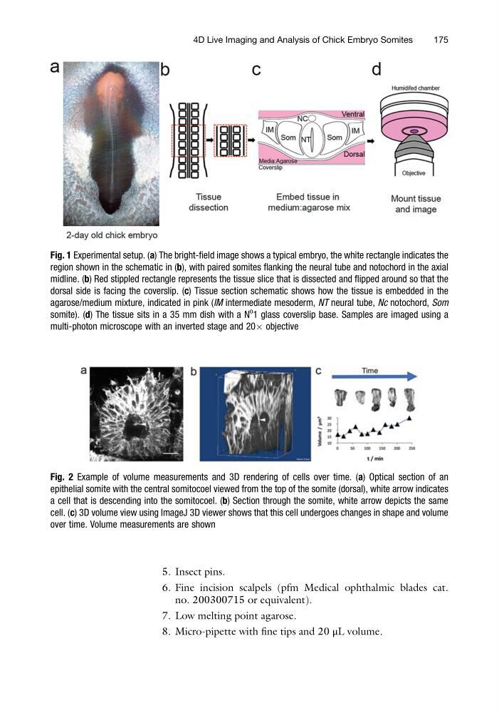
Fig. 1 Experimental setup. (a) The bright-field image shows a typical embryo, the white rectangle indicates the
region shown in the schematic in (b), with paired somites flanking the neural tube and notochord in the axial
midline. (b) Red stippled rectangle represents the tissue slice that is dissected and flipped around so that the
dorsal side is facing the coverslip. (c) Tissue section schematic shows how the tissue is embedded in the
agarose/medium mixture, indicated in pink (IM intermediate mesoderm, NT neural tube, Nc notochord, Som
somite). (d) The tissue sits in a 35 mm dish with a N
o 1 glass coverslip base. Samples are imaged using a
multi-photon microscope with an inverted stage and 20Â objective
Fig. 2 Example of volume measurements and 3D rendering of cells over time. (a) Optical section of an
epithelial somite with the central somitocoel viewed from the top of the somite (dorsal), white arrow indicates
a cell that is descending into the somitocoel. (b) Section through the somite, white arrow depicts the same
cell. (c) 3D volume view using ImageJ 3D viewer shows that this cell undergoes changes in shape and volume
over time. Volume measurements are shown
4D Live Imaging and Analysis of Chick Embryo Somites
175
6. Fine incision scalpels (pfm Medical ophthalmic blades cat.
no. 200300715 or equivalent).
7. Low melting point agarose.
8. Micro-pipette with fine tips and 20 μL volume.
Fig. 1 Experimental setup. (a) The bright-field image shows a typical embryo, the white rectangle indicates the
region shown in the schematic in (b), with paired somites flanking the neural tube and notochord in the axial
midline. (b) Red stippled rectangle represents the tissue slice that is dissected and flipped around so that the
dorsal side is facing the coverslip. (c) Tissue section schematic shows how the tissue is embedded in the
agarose/medium mixture, indicated in pink (IM intermediate mesoderm, NT neural tube, Nc notochord, Som
somite). (d) The tissue sits in a 35 mm dish with a N
o 1 glass coverslip base. Samples are imaged using a
multi-photon microscope with an inverted stage and 20Â objective
Fig. 2 Example of volume measurements and 3D rendering of cells over time. (a) Optical section of an
epithelial somite with the central somitocoel viewed from the top of the somite (dorsal), white arrow indicates
a cell that is descending into the somitocoel. (b) Section through the somite, white arrow depicts the same
cell. (c) 3D volume view using ImageJ 3D viewer shows that this cell undergoes changes in shape and volume
over time. Volume measurements are shown
4D Live Imaging and Analysis of Chick Embryo Somites
175