

2. After sacrificing the female, dissect out the uterus (remove as
much fat as possible) and place it in a dish of dissection
medium.
3. Under a stereomicroscope, dissect deciduae out of the uterus.
Make a slit on the decidua side. When the slit is almost as
important as the height of the decidua, make a slit on the side
of one decidua and pull out the decidua by the opening. It is
best to use coarse forceps at this stage.
4. Place deciduae in a dish with clean medium to improve visibility. Deciduae are pear shaped, and embryos are within their
broader section. Hold the broad extremity with one hand, and
slice the thinner portion in 2 using not too sharp forceps, in
order to be able to open the decidua as an open book. Carefully
remove embryos from the deciduae with closed forceps by
digging under the embryo and gently pushing it.
5. Place embryos in yet another dish with clean medium. Remove
Reichert’s membrane with either fine pristine forceps or sharpened tungsten needles. It is essential that embryo and ectoplacental cone are left intact, and damaged embryos should not be
used for imaging. Transfer the embryo into culture medium with
a pipette or tip so as to transfer a minimal amount of dissection
medium and place in the incubator (see Notes 4 and 5).
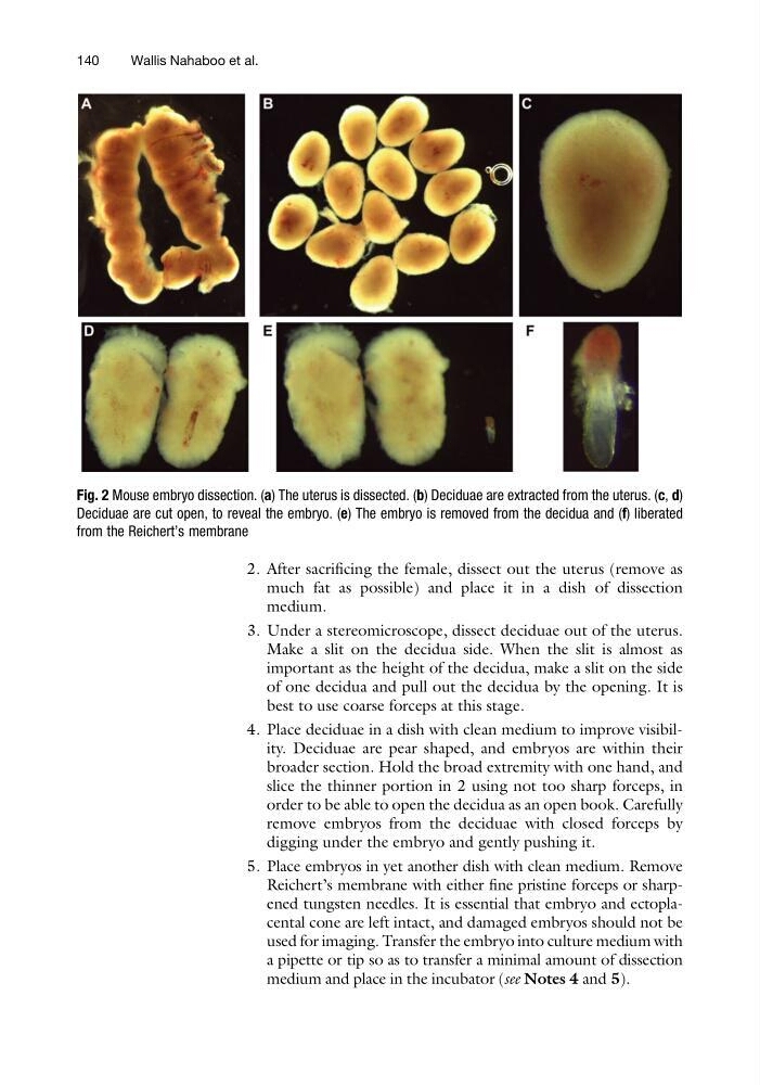
Fig. 2 Mouse embryo dissection. (a) The uterus is dissected. (b) Deciduae are extracted from the uterus. (c, d)
Deciduae are cut open, to reveal the embryo. (e) The embryo is removed from the decidua and (f) liberated
from the Reichert’s membrane
140
Wallis Nahaboo et al.
much fat as possible) and place it in a dish of dissection
medium.
3. Under a stereomicroscope, dissect deciduae out of the uterus.
Make a slit on the decidua side. When the slit is almost as
important as the height of the decidua, make a slit on the side
of one decidua and pull out the decidua by the opening. It is
best to use coarse forceps at this stage.
4. Place deciduae in a dish with clean medium to improve visibility. Deciduae are pear shaped, and embryos are within their
broader section. Hold the broad extremity with one hand, and
slice the thinner portion in 2 using not too sharp forceps, in
order to be able to open the decidua as an open book. Carefully
remove embryos from the deciduae with closed forceps by
digging under the embryo and gently pushing it.
5. Place embryos in yet another dish with clean medium. Remove
Reichert’s membrane with either fine pristine forceps or sharpened tungsten needles. It is essential that embryo and ectoplacental cone are left intact, and damaged embryos should not be
used for imaging. Transfer the embryo into culture medium with
a pipette or tip so as to transfer a minimal amount of dissection
medium and place in the incubator (see Notes 4 and 5).
Fig. 2 Mouse embryo dissection. (a) The uterus is dissected. (b) Deciduae are extracted from the uterus. (c, d)
Deciduae are cut open, to reveal the embryo. (e) The embryo is removed from the decidua and (f) liberated
from the Reichert’s membrane
140
Wallis Nahaboo et al.