

over the area to be imaged with a tungsten needle or forceps
and tack down by sticking it to the filter paper. Let albumen run
over paper while in the egg and moisten any dry parts with
Ringer’s solution.
4. Cut around outside of paper ring to remove embryo. Do not
cut any vasculature. Depending on the developmental stage,
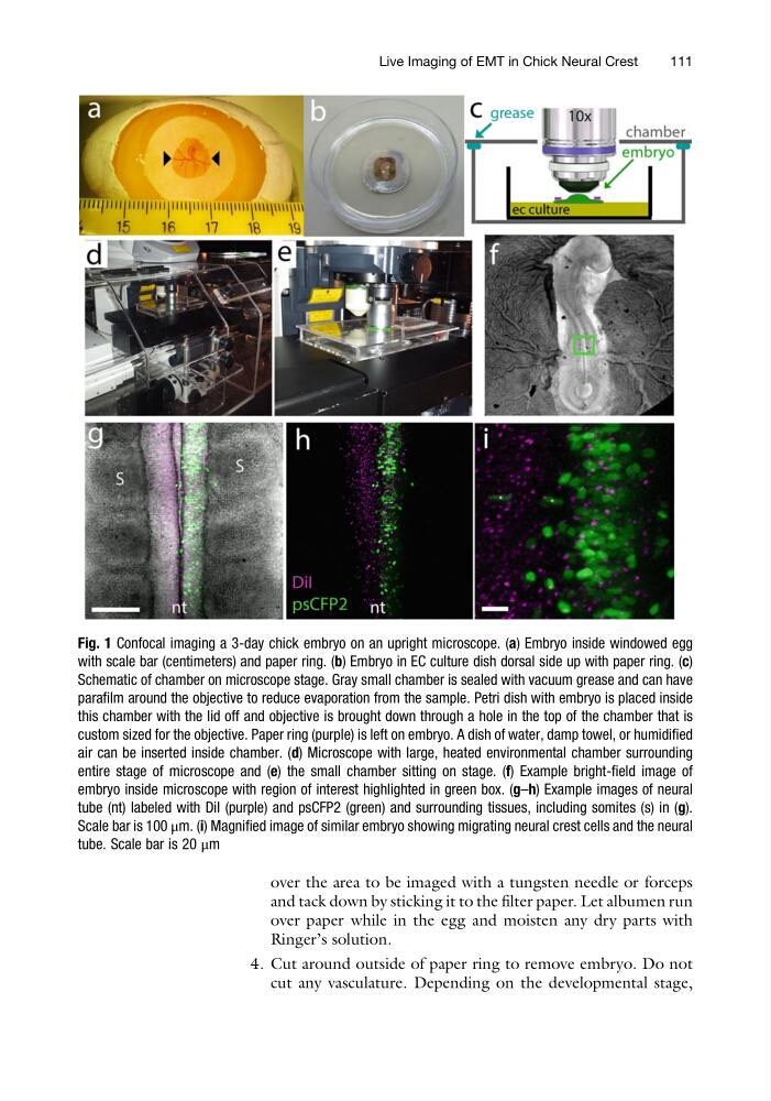
Fig. 1 Confocal imaging a 3-day chick embryo on an upright microscope. (a) Embryo inside windowed egg
with scale bar (centimeters) and paper ring. (b) Embryo in EC culture dish dorsal side up with paper ring. (c)
Schematic of chamber on microscope stage. Gray small chamber is sealed with vacuum grease and can have
parafilm around the objective to reduce evaporation from the sample. Petri dish with embryo is placed inside
this chamber with the lid off and objective is brought down through a hole in the top of the chamber that is
custom sized for the objective. Paper ring (purple) is left on embryo. A dish of water, damp towel, or humidified
air can be inserted inside chamber. (d) Microscope with large, heated environmental chamber surrounding
entire stage of microscope and (e) the small chamber sitting on stage. (f) Example bright-field image of
embryo inside microscope with region of interest highlighted in green box. (g–h) Example images of neural
tube (nt) labeled with DiI (purple) and psCFP2 (green) and surrounding tissues, including somites (s) in (g).
Scale bar is 100 μm. (i) Magnified image of similar embryo showing migrating neural crest cells and the neural
tube. Scale bar is 20 μm
Live Imaging of EMT in Chick Neural Crest
111
and tack down by sticking it to the filter paper. Let albumen run
over paper while in the egg and moisten any dry parts with
Ringer’s solution.
4. Cut around outside of paper ring to remove embryo. Do not
cut any vasculature. Depending on the developmental stage,
Fig. 1 Confocal imaging a 3-day chick embryo on an upright microscope. (a) Embryo inside windowed egg
with scale bar (centimeters) and paper ring. (b) Embryo in EC culture dish dorsal side up with paper ring. (c)
Schematic of chamber on microscope stage. Gray small chamber is sealed with vacuum grease and can have
parafilm around the objective to reduce evaporation from the sample. Petri dish with embryo is placed inside
this chamber with the lid off and objective is brought down through a hole in the top of the chamber that is
custom sized for the objective. Paper ring (purple) is left on embryo. A dish of water, damp towel, or humidified
air can be inserted inside chamber. (d) Microscope with large, heated environmental chamber surrounding
entire stage of microscope and (e) the small chamber sitting on stage. (f) Example bright-field image of
embryo inside microscope with region of interest highlighted in green box. (g–h) Example images of neural
tube (nt) labeled with DiI (purple) and psCFP2 (green) and surrounding tissues, including somites (s) in (g).
Scale bar is 100 μm. (i) Magnified image of similar embryo showing migrating neural crest cells and the neural
tube. Scale bar is 20 μm
Live Imaging of EMT in Chick Neural Crest
111