

30
L. Quadro and E. K. Spiegler
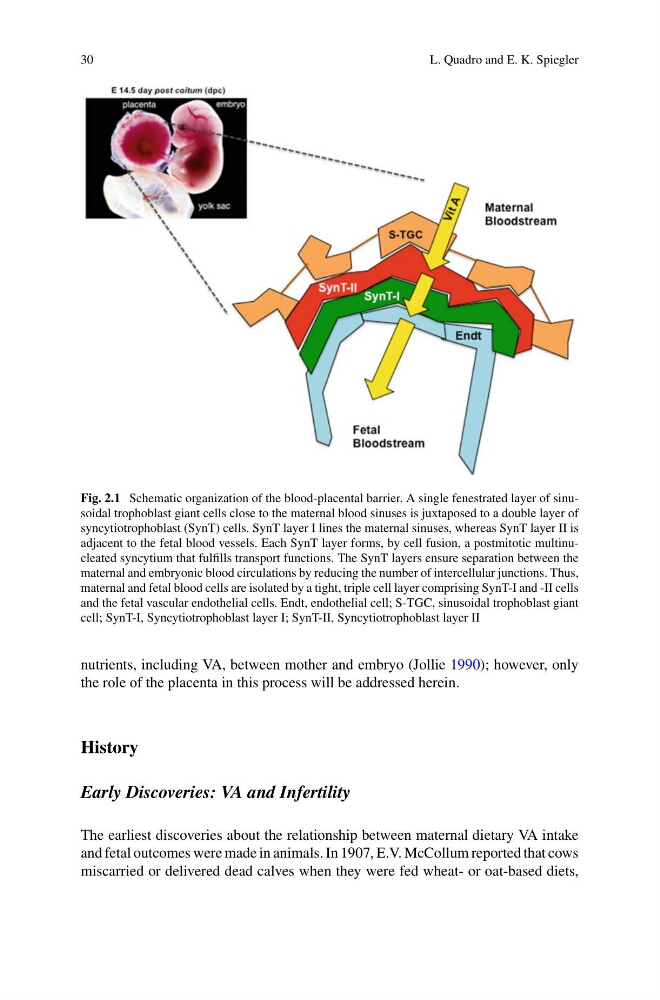
Fig. 2.1 Schematic organization of the blood-placental barrier. A single fenestrated layer of sinusoidal trophoblast giant cells close to the maternal blood sinuses is juxtaposed to a double layer of
syncytiotrophoblast (SynT) cells. SynT layer I lines the maternal sinuses, whereas SynT layer II is
adjacent to the fetal blood vessels. Each SynT layer forms, by cell fusion, a postmitotic multinucleated syncytium that fulfills transport functions. The SynT layers ensure separation between the
maternal and embryonic blood circulations by reducing the number of intercellular junctions. Thus,
maternal and fetal blood cells are isolated by a tight, triple cell layer comprising SynT-I and -II cells
and the fetal vascular endothelial cells. Endt, endothelial cell; S-TGC, sinusoidal trophoblast giant
cell; SynT-I, Syncytiotrophoblast layer I; SynT-II, Syncytiotrophoblast layer II
nutrients, including VA, between mother and embryo (Jollie 1990); however, only
the role of the placenta in this process will be addressed herein.
History
Early Discoveries: VA and Infertility
The earliest discoveries about the relationship between maternal dietary VA intake
and fetal outcomes were made in animals. In 1907, E.V. McCollum reported that cows
miscarried or delivered dead calves when they were fed wheat- or oat-based diets,
L. Quadro and E. K. Spiegler
Fig. 2.1 Schematic organization of the blood-placental barrier. A single fenestrated layer of sinusoidal trophoblast giant cells close to the maternal blood sinuses is juxtaposed to a double layer of
syncytiotrophoblast (SynT) cells. SynT layer I lines the maternal sinuses, whereas SynT layer II is
adjacent to the fetal blood vessels. Each SynT layer forms, by cell fusion, a postmitotic multinucleated syncytium that fulfills transport functions. The SynT layers ensure separation between the
maternal and embryonic blood circulations by reducing the number of intercellular junctions. Thus,
maternal and fetal blood cells are isolated by a tight, triple cell layer comprising SynT-I and -II cells
and the fetal vascular endothelial cells. Endt, endothelial cell; S-TGC, sinusoidal trophoblast giant
cell; SynT-I, Syncytiotrophoblast layer I; SynT-II, Syncytiotrophoblast layer II
nutrients, including VA, between mother and embryo (Jollie 1990); however, only
the role of the placenta in this process will be addressed herein.
History
Early Discoveries: VA and Infertility
The earliest discoveries about the relationship between maternal dietary VA intake
and fetal outcomes were made in animals. In 1907, E.V. McCollum reported that cows
miscarried or delivered dead calves when they were fed wheat- or oat-based diets,