

16
M. A. Asson-Batres
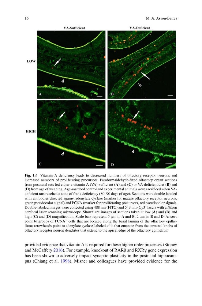
Fig. 1.4 Vitamin A deficiency leads to decreased numbers of olfactory receptor neurons and
increased numbers of proliferating precursors. Paraformaldehyde-fixed olfactory organ sections
from postnatal rats fed either a vitamin A (VA)-sufficient (A) and (C) or VA-deficient diet (B) and
(D) from age of weaning. Age-matched control and experimental animals were sacrificed when VAdeficient rats reached a state of frank deficiency (80–90 days of age). Sections were double labeled
with antibodies directed against adenylate cyclase (marker for mature olfactory receptor neurons,
green pseudocolor signal) and PCNA (marker for proliferating precursors, red pseudocolor signal).
Double-labeled images were collected using 488 nm (FITC) and 543 nm (Cy3) lasers with a Nikon
confocal laser scanning microscope. Shown are images of sections taken at low (A) and (B) and
high (C) and (D) magnification. Scale bars represent 5 μm in A and B, 2 μm in B and D. Arrows
point to groups of PCNA + cells that are located along the basal lamina of the olfactory epithelium; arrowheads point to adenylate cyclase-labeled cilia that emanate from the terminal knobs of
olfactory receptor neuron dendrites that extend to the apical edge of the olfactory epithelium
provided evidence that vitamin A is required for these higher order processes (Stoney
and McCaffery 2016). For example, knockout of RARβ and RXRγ gene expression
has been shown to adversely impact synaptic plasticity in the postnatal hippocampus (Chiang et al. 1998). Misner and colleagues have provided evidence for the
M. A. Asson-Batres
Fig. 1.4 Vitamin A deficiency leads to decreased numbers of olfactory receptor neurons and
increased numbers of proliferating precursors. Paraformaldehyde-fixed olfactory organ sections
from postnatal rats fed either a vitamin A (VA)-sufficient (A) and (C) or VA-deficient diet (B) and
(D) from age of weaning. Age-matched control and experimental animals were sacrificed when VAdeficient rats reached a state of frank deficiency (80–90 days of age). Sections were double labeled
with antibodies directed against adenylate cyclase (marker for mature olfactory receptor neurons,
green pseudocolor signal) and PCNA (marker for proliferating precursors, red pseudocolor signal).
Double-labeled images were collected using 488 nm (FITC) and 543 nm (Cy3) lasers with a Nikon
confocal laser scanning microscope. Shown are images of sections taken at low (A) and (B) and
high (C) and (D) magnification. Scale bars represent 5 μm in A and B, 2 μm in B and D. Arrows
point to groups of PCNA + cells that are located along the basal lamina of the olfactory epithelium; arrowheads point to adenylate cyclase-labeled cilia that emanate from the terminal knobs of
olfactory receptor neuron dendrites that extend to the apical edge of the olfactory epithelium
provided evidence that vitamin A is required for these higher order processes (Stoney
and McCaffery 2016). For example, knockout of RARβ and RXRγ gene expression
has been shown to adversely impact synaptic plasticity in the postnatal hippocampus (Chiang et al. 1998). Misner and colleagues have provided evidence for the