

188
W. R. Jackman and Y. Gibert
are not able to form due to indirect effects. Later RA inhibition during gastrulation
is likely altering CNC cell specification, which as a result could be depleting the
tooth-forming region of odontoblast precursors. However, because no morphological sign of tooth development, or even early tooth-prefiguring gene expression, can
be seen in each of these sets of relatively early RA inhibition experiments, it is difficult to speculate in a more specific way what the function of RA during early stages
of tooth development may be. In contrast, later RA inhibition at about 40 hpf also
completely prevents morphological evidence of tooth development, but some very
early tooth-related gene expression is at least party retained.
The earliest known gene to be expressed specifically during tooth formation is the
transcription factor pitx2, which is activated in cells of the pharyngeal epithelium at
the location of tooth formation, including some cells that will later likely become
ameloblasts (Jackman et al. 2004). When RA signaling is inhibited starting at 40
hpf, some pitx2 expression remains localized to the tooth forming region, but in a
disrupted pattern (Gibert et al. 2010). From this gene expression result, it can be
concluded that RA is likely not the earliest cue to initiate normal tooth formation,
but that it does play a necessary role very soon after tooth-specific gene expression
commences. Because later RA inhibition has no effect on tooth formation, it is also
reasonable to conclude that RA does not normally participate in any later stage of
tooth morphogenesis or mineralization. This is somewhat different than the effects on
mineralization that were observed classically in the continuously developing rodent
incisor, and this difference may reflect a new role that RA has acquired in the evolution
of the unusual development of an ever-growing tooth type in rodents.
Regarding the 24 hpf exogenous RA experiments in zebrafish, perhaps the two
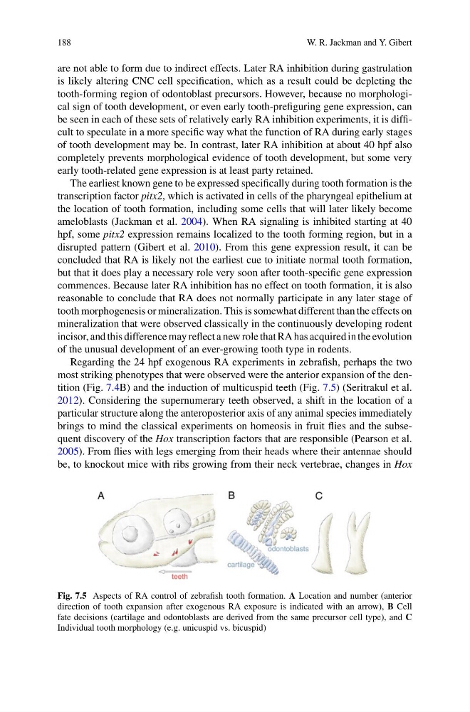
most striking phenotypes that were observed were the anterior expansion of the dentition (Fig. 7.4B) and the induction of multicuspid teeth (Fig. 7.5) (Seritrakul et al.
2012). Considering the supernumerary teeth observed, a shift in the location of a
particular structure along the anteroposterior axis of any animal species immediately
brings to mind the classical experiments on homeosis in fruit flies and the subsequent discovery of the Hox transcription factors that are responsible (Pearson et al.
2005). From flies with legs emerging from their heads where their antennae should
be, to knockout mice with ribs growing from their neck vertebrae, changes in Hox
Fig. 7.5 Aspects of RA control of zebrafish tooth formation. A Location and number (anterior
direction of tooth expansion after exogenous RA exposure is indicated with an arrow), B Cell
fate decisions (cartilage and odontoblasts are derived from the same precursor cell type), and C
Individual tooth morphology (e.g. unicuspid vs. bicuspid)
W. R. Jackman and Y. Gibert
are not able to form due to indirect effects. Later RA inhibition during gastrulation
is likely altering CNC cell specification, which as a result could be depleting the
tooth-forming region of odontoblast precursors. However, because no morphological sign of tooth development, or even early tooth-prefiguring gene expression, can
be seen in each of these sets of relatively early RA inhibition experiments, it is difficult to speculate in a more specific way what the function of RA during early stages
of tooth development may be. In contrast, later RA inhibition at about 40 hpf also
completely prevents morphological evidence of tooth development, but some very
early tooth-related gene expression is at least party retained.
The earliest known gene to be expressed specifically during tooth formation is the
transcription factor pitx2, which is activated in cells of the pharyngeal epithelium at
the location of tooth formation, including some cells that will later likely become
ameloblasts (Jackman et al. 2004). When RA signaling is inhibited starting at 40
hpf, some pitx2 expression remains localized to the tooth forming region, but in a
disrupted pattern (Gibert et al. 2010). From this gene expression result, it can be
concluded that RA is likely not the earliest cue to initiate normal tooth formation,
but that it does play a necessary role very soon after tooth-specific gene expression
commences. Because later RA inhibition has no effect on tooth formation, it is also
reasonable to conclude that RA does not normally participate in any later stage of
tooth morphogenesis or mineralization. This is somewhat different than the effects on
mineralization that were observed classically in the continuously developing rodent
incisor, and this difference may reflect a new role that RA has acquired in the evolution
of the unusual development of an ever-growing tooth type in rodents.
Regarding the 24 hpf exogenous RA experiments in zebrafish, perhaps the two
most striking phenotypes that were observed were the anterior expansion of the dentition (Fig. 7.4B) and the induction of multicuspid teeth (Fig. 7.5) (Seritrakul et al.
2012). Considering the supernumerary teeth observed, a shift in the location of a
particular structure along the anteroposterior axis of any animal species immediately
brings to mind the classical experiments on homeosis in fruit flies and the subsequent discovery of the Hox transcription factors that are responsible (Pearson et al.
2005). From flies with legs emerging from their heads where their antennae should
be, to knockout mice with ribs growing from their neck vertebrae, changes in Hox
Fig. 7.5 Aspects of RA control of zebrafish tooth formation. A Location and number (anterior
direction of tooth expansion after exogenous RA exposure is indicated with an arrow), B Cell
fate decisions (cartilage and odontoblasts are derived from the same precursor cell type), and C
Individual tooth morphology (e.g. unicuspid vs. bicuspid)