

156
H. A. Marquez and F. Chen
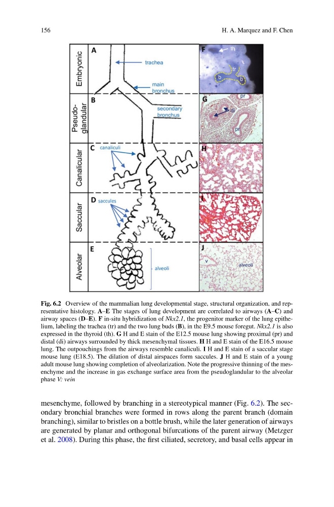
Fig. 6.2 Overview of the mammalian lung developmental stage, structural organization, and representative histology. A–E The stages of lung development are correlated to airways (A–C) and
airway spaces (D–E). F in-situ hybridization of Nkx2.1, the progenitor marker of the lung epithelium, labeling the trachea (tr) and the two lung buds (B), in the E9.5 mouse foregut. Nkx2.1 is also
expressed in the thyroid (th). G H and E stain of the E12.5 mouse lung showing proximal (pr) and
distal (di) airways surrounded by thick mesenchymal tissues. H H and E stain of the E16.5 mouse
lung. The outpouchings from the airways resemble canaliculi. I H and E stain of a saccular stage
mouse lung (E18.5). The dilation of distal airspaces form saccules. J H and E stain of a young
adult mouse lung showing completion of alveolarization. Note the progressive thinning of the mesenchyme and the increase in gas exchange surface area from the pseudoglandular to the alveolar
phase V: vein
mesenchyme, followed by branching in a stereotypical manner (Fig. 6.2). The secondary bronchial branches were formed in rows along the parent branch (domain
branching), similar to bristles on a bottle brush, while the later generation of airways
are generated by planar and orthogonal bifurcations of the parent airway (Metzger
et al. 2008). During this phase, the first ciliated, secretory, and basal cells appear in
H. A. Marquez and F. Chen
Fig. 6.2 Overview of the mammalian lung developmental stage, structural organization, and representative histology. A–E The stages of lung development are correlated to airways (A–C) and
airway spaces (D–E). F in-situ hybridization of Nkx2.1, the progenitor marker of the lung epithelium, labeling the trachea (tr) and the two lung buds (B), in the E9.5 mouse foregut. Nkx2.1 is also
expressed in the thyroid (th). G H and E stain of the E12.5 mouse lung showing proximal (pr) and
distal (di) airways surrounded by thick mesenchymal tissues. H H and E stain of the E16.5 mouse
lung. The outpouchings from the airways resemble canaliculi. I H and E stain of a saccular stage
mouse lung (E18.5). The dilation of distal airspaces form saccules. J H and E stain of a young
adult mouse lung showing completion of alveolarization. Note the progressive thinning of the mesenchyme and the increase in gas exchange surface area from the pseudoglandular to the alveolar
phase V: vein
mesenchyme, followed by branching in a stereotypical manner (Fig. 6.2). The secondary bronchial branches were formed in rows along the parent branch (domain
branching), similar to bristles on a bottle brush, while the later generation of airways
are generated by planar and orthogonal bifurcations of the parent airway (Metzger
et al. 2008). During this phase, the first ciliated, secretory, and basal cells appear in