

5 Retinoic Acid Signaling and Heart Development
123
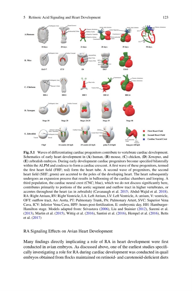
Fig. 5.1 Waves of differentiating cardiac progenitors contribute to vertebrate cardiac development.
Schematics of early heart development in (A) human, (B) mouse, (C) chicken, (D) Xenopus, and
(E) zebrafish embryos. During early development cardiac progenitors become specified bilaterally
within the ALPM and coalesce to form a cardiac crescent. A first wave of these progenitors, termed
the first heart field (FHF; red) form the heart tube. A second wave of progenitors, the second
heart field (SHF; green) are accreted to the poles of the developing heart. The heart subsequently
undergoes an expansion process that results in ballooning of the cardiac chambers and looping. A
third population, the cardiac neural crest (CNC; blue), which we do not discuss significantly here,
contributes primarily to portions of the aortic segment and outflow tract in higher vertebrates, or
accretes throughout the heart (as in zebrafish) (Cavanaugh et al. 2015; Abdul-Wajid et al. 2018).
RA: Right Atrium, RV: Right Ventricle, LA: Left Atrium, LV: Left Ventricle, A: atrium, V: ventricle,
OFT: outflow tract, Ao: Aorta, PT: Pulmonary Trunk, PA: Pulmonary Artert, SVC: Superior Vena
Cava, ICV: Inferior Vena Cava, HPF: hours post-fertilization, E: embryonic day, HH: HamburgerHamilton stage. Models adapted from: Srivastava (2006), Liu and Stainier (2012), Saremi et al.
(2013), Martin et al. (2015), Wittig et al. (2016), Santini et al. (2016), Hempel et al. (2016), Betts
et al. (2017)
RA Signaling Effects on Avian Heart Development
Many findings directly implicating a role of RA in heart development were first
conducted in avian embryos. As discussed above, one of the earliest studies specifically investigating a role for RA during cardiac development was conducted in quail
embryos obtained from flocks maintained on retinoid- and carotenoid-deficient diets
123
Fig. 5.1 Waves of differentiating cardiac progenitors contribute to vertebrate cardiac development.
Schematics of early heart development in (A) human, (B) mouse, (C) chicken, (D) Xenopus, and
(E) zebrafish embryos. During early development cardiac progenitors become specified bilaterally
within the ALPM and coalesce to form a cardiac crescent. A first wave of these progenitors, termed
the first heart field (FHF; red) form the heart tube. A second wave of progenitors, the second
heart field (SHF; green) are accreted to the poles of the developing heart. The heart subsequently
undergoes an expansion process that results in ballooning of the cardiac chambers and looping. A
third population, the cardiac neural crest (CNC; blue), which we do not discuss significantly here,
contributes primarily to portions of the aortic segment and outflow tract in higher vertebrates, or
accretes throughout the heart (as in zebrafish) (Cavanaugh et al. 2015; Abdul-Wajid et al. 2018).
RA: Right Atrium, RV: Right Ventricle, LA: Left Atrium, LV: Left Ventricle, A: atrium, V: ventricle,
OFT: outflow tract, Ao: Aorta, PT: Pulmonary Trunk, PA: Pulmonary Artert, SVC: Superior Vena
Cava, ICV: Inferior Vena Cava, HPF: hours post-fertilization, E: embryonic day, HH: HamburgerHamilton stage. Models adapted from: Srivastava (2006), Liu and Stainier (2012), Saremi et al.
(2013), Martin et al. (2015), Wittig et al. (2016), Santini et al. (2016), Hempel et al. (2016), Betts
et al. (2017)
RA Signaling Effects on Avian Heart Development
Many findings directly implicating a role of RA in heart development were first
conducted in avian embryos. As discussed above, one of the earliest studies specifically investigating a role for RA during cardiac development was conducted in quail
embryos obtained from flocks maintained on retinoid- and carotenoid-deficient diets