

79
antioxidant properties. Cell proliferation increased on the PCL/lignin-PCL nanofibrous scaffolds, especially under cellular oxidative stress. In addition, nanofibrous
scaffolds improved the neurite outgrowth of DRG neurons and myelin basic protein
expressions of Schwann cells. PCL, as well as poly(lactic acid) (PLA), are also
commonly used as biomaterials for tissue engineering. Kai et al. (2016b) incorporated lignin into PLA to defend against oxidative stress induced by PLA as a biomaterial. Poly(L-lactic acid) (PLLA)/PLA-lignin nanofibers were prepared by
electrospinning. Three types of cells cultured on the PLLA/PLA-lignin nanofibers,
such as rat pheochromyctoma (PC-12) cells, hMSCs and human dermal fibroblasts
(HDFs), exhibited a higher proliferation rate than those on pure PLLA nanofibers.
This is because the antioxidant activity of the PLLA/PLA-lignin nanofibers
improves the viability of the cells, but to obtain the best biocompatibility with the
cell, a balance between cytotoxicity and antioxidant activities must be achieved by
optimizing the amount of lignin in PLLA/PLA-lignin nanofibers. Kai et al. (2017)
prepared PLLA/lignin-poly(ε-caprolactone-co-lactide) and poly(ε-caprolactone)/
lignin-poly(ε-caprolactone-co-lactide) nanofibers. When cultured on these two
nanofibers, the NIH/3 T3 fibroblasts were well distributed and oriented along fibers.
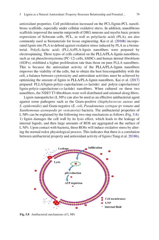
Lignin nanoparticles (L NPs) can also be used as an effective antibacterial agent
against some pathogens such as the Gram-positive (Staphylococcus aureus and
S. epidermidis) and Gram-negative (E. coli, Pseudomonas syringae pv tomato and
Xanthomonas axonopodis pv vesicatoria) bacteria. The antibacterial properties of
L NPs can be explained by the following two-step mechanism as follows (Fig. 5.8):
1) lignin damages the cell wall by its lysis effect, which leads to the leakage of
internal liquids, and then large amounts of ROS are aggregated on the surface of
L NPs. Upon contact with bacteria, these ROSs will induce oxidative stress by altering the normal redox physiological process. This indicates that there is a correlation
between antibacterial property and antioxidant activity of lignin (Yang et al. 2018b),
LNP
Cell membrance
Internal fluid
pH reduction
ATP depletion
Lysis
Leakage
Infiltration
Fig. 5.8 Antibacterial mechanisms of L NPs
5 Lignin as a Natural Antioxidant: Property-Structure Relationship and Potential…
antioxidant properties. Cell proliferation increased on the PCL/lignin-PCL nanofibrous scaffolds, especially under cellular oxidative stress. In addition, nanofibrous
scaffolds improved the neurite outgrowth of DRG neurons and myelin basic protein
expressions of Schwann cells. PCL, as well as poly(lactic acid) (PLA), are also
commonly used as biomaterials for tissue engineering. Kai et al. (2016b) incorporated lignin into PLA to defend against oxidative stress induced by PLA as a biomaterial. Poly(L-lactic acid) (PLLA)/PLA-lignin nanofibers were prepared by
electrospinning. Three types of cells cultured on the PLLA/PLA-lignin nanofibers,
such as rat pheochromyctoma (PC-12) cells, hMSCs and human dermal fibroblasts
(HDFs), exhibited a higher proliferation rate than those on pure PLLA nanofibers.
This is because the antioxidant activity of the PLLA/PLA-lignin nanofibers
improves the viability of the cells, but to obtain the best biocompatibility with the
cell, a balance between cytotoxicity and antioxidant activities must be achieved by
optimizing the amount of lignin in PLLA/PLA-lignin nanofibers. Kai et al. (2017)
prepared PLLA/lignin-poly(ε-caprolactone-co-lactide) and poly(ε-caprolactone)/
lignin-poly(ε-caprolactone-co-lactide) nanofibers. When cultured on these two
nanofibers, the NIH/3 T3 fibroblasts were well distributed and oriented along fibers.
Lignin nanoparticles (L NPs) can also be used as an effective antibacterial agent
against some pathogens such as the Gram-positive (Staphylococcus aureus and
S. epidermidis) and Gram-negative (E. coli, Pseudomonas syringae pv tomato and
Xanthomonas axonopodis pv vesicatoria) bacteria. The antibacterial properties of
L NPs can be explained by the following two-step mechanism as follows (Fig. 5.8):
1) lignin damages the cell wall by its lysis effect, which leads to the leakage of
internal liquids, and then large amounts of ROS are aggregated on the surface of
L NPs. Upon contact with bacteria, these ROSs will induce oxidative stress by altering the normal redox physiological process. This indicates that there is a correlation
between antibacterial property and antioxidant activity of lignin (Yang et al. 2018b),
LNP
Cell membrance
Internal fluid
pH reduction
ATP depletion
Lysis
Leakage
Infiltration
Fig. 5.8 Antibacterial mechanisms of L NPs
5 Lignin as a Natural Antioxidant: Property-Structure Relationship and Potential…