

3.3 Label-Free
Quantitative Proteomic
Analysis: 1-D
SDS-PAGE
1. Dilute the protein sample if needed with ultrapure water and
mix the same quantity of total protein from each individual
sample (about 10–20 μg) with loading buffer in a 2:1 protein
extract:buffer volume ratio.
2. Heat samples in thermoblock at 100
C for 5 min. Centrifuge
1 min at 4000 x g.
3. Load samples (do not exceed the maximum volume; follow the
guidelines indicated in the instruction manual of Mini-PROTEAN
® Tetra Vertical Electrophoresis Cell, Bio-Rad Laboratories, USA) in each lane of an SDS-polyacrylamide gel of
1 mm thickness (12.5%, including a top layer of 4%-stacking
gel) (see Note 5).
4. Proteins are separated electrophoretically based on classical
SDS-PAGE Laemmli Buffer System according to molecular
weight (MW) using Tris-glycine-SDS running buffer at
180 V, 15 min (to get enough separation to divide the samples
in three fractions of different MW) or using longer times/
different voltages depending on the specific user needs (see
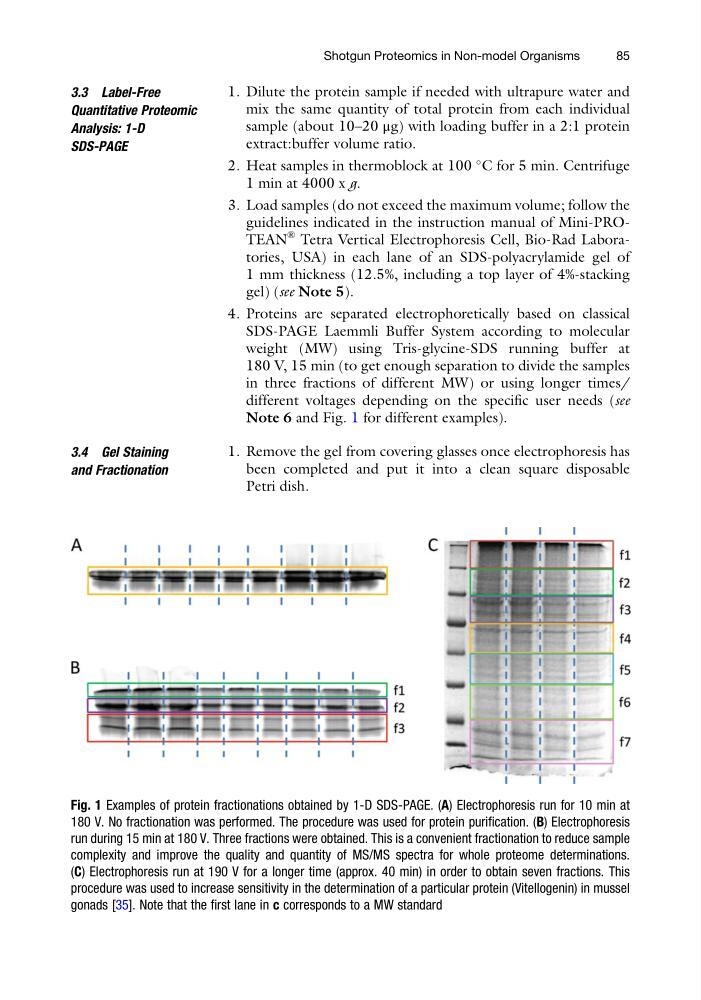
Note 6 and Fig. 1 for different examples).
3.4 Gel Staining
and Fractionation
1. Remove the gel from covering glasses once electrophoresis has
been completed and put it into a clean square disposable
Petri dish.
Fig. 1 Examples of protein fractionations obtained by 1-D SDS-PAGE. (A) Electrophoresis run for 10 min at
180 V. No fractionation was performed. The procedure was used for protein purification. (B) Electrophoresis
run during 15 min at 180 V. Three fractions were obtained. This is a convenient fractionation to reduce sample
complexity and improve the quality and quantity of MS/MS spectra for whole proteome determinations.
(C) Electrophoresis run at 190 V for a longer time (approx. 40 min) in order to obtain seven fractions. This
procedure was used to increase sensitivity in the determination of a particular protein (Vitellogenin) in mussel
gonads [35]. Note that the first lane in c corresponds to a MW standard
Shotgun Proteomics in Non-model Organisms
85
Quantitative Proteomic
Analysis: 1-D
SDS-PAGE
1. Dilute the protein sample if needed with ultrapure water and
mix the same quantity of total protein from each individual
sample (about 10–20 μg) with loading buffer in a 2:1 protein
extract:buffer volume ratio.
2. Heat samples in thermoblock at 100
C for 5 min. Centrifuge
1 min at 4000 x g.
3. Load samples (do not exceed the maximum volume; follow the
guidelines indicated in the instruction manual of Mini-PROTEAN
® Tetra Vertical Electrophoresis Cell, Bio-Rad Laboratories, USA) in each lane of an SDS-polyacrylamide gel of
1 mm thickness (12.5%, including a top layer of 4%-stacking
gel) (see Note 5).
4. Proteins are separated electrophoretically based on classical
SDS-PAGE Laemmli Buffer System according to molecular
weight (MW) using Tris-glycine-SDS running buffer at
180 V, 15 min (to get enough separation to divide the samples
in three fractions of different MW) or using longer times/
different voltages depending on the specific user needs (see
Note 6 and Fig. 1 for different examples).
3.4 Gel Staining
and Fractionation
1. Remove the gel from covering glasses once electrophoresis has
been completed and put it into a clean square disposable
Petri dish.
Fig. 1 Examples of protein fractionations obtained by 1-D SDS-PAGE. (A) Electrophoresis run for 10 min at
180 V. No fractionation was performed. The procedure was used for protein purification. (B) Electrophoresis
run during 15 min at 180 V. Three fractions were obtained. This is a convenient fractionation to reduce sample
complexity and improve the quality and quantity of MS/MS spectra for whole proteome determinations.
(C) Electrophoresis run at 190 V for a longer time (approx. 40 min) in order to obtain seven fractions. This
procedure was used to increase sensitivity in the determination of a particular protein (Vitellogenin) in mussel
gonads [35]. Note that the first lane in c corresponds to a MW standard
Shotgun Proteomics in Non-model Organisms
85