

8. After 1 week, collect the L4 stage larvae and go to protein
extraction and precipitation (see Subheading 3.2).
3.2 Protein
Extraction
and Precipitation
1. Homogenize mechanically on ice the L3 and L4 larvae in 2 mL
centrifuge tubes with an addition of 1.5 mL of lysis buffer per
sample. Prepare five samples of L3 development stage by
homogenizing 5–10 larvae in each sample and next five samples
of L4 development stage by homogenizing 5–10 larvae in each
sample (see Note 11).
2. Sonicate the samples on ice for 3 cycles of 30 s pulses (see
Note 12).
3. Centrifuge lysate at 16,000 Â g for 30 min at 4
C.
4. Carefully separate the supernatant and transfer into a new tube.
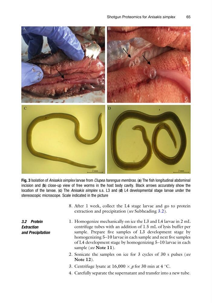
Fig. 3 Isolation of Anisakis simplex larvae from Clupea harengus membras. (a) The fish longitudinal abdominal
incision and (b) close-up view of free worms in the host body cavity. Black arrows accurately show the
location of the larvae. (c) The Anisakis simplex s.s. L3 and (d) L4 developmental stage larvae under the
stereoscopic microscope. Scale indicated in the picture
Shotgun Proteomics for Anisakis simplex
65
extraction and precipitation (see Subheading 3.2).
3.2 Protein
Extraction
and Precipitation
1. Homogenize mechanically on ice the L3 and L4 larvae in 2 mL
centrifuge tubes with an addition of 1.5 mL of lysis buffer per
sample. Prepare five samples of L3 development stage by
homogenizing 5–10 larvae in each sample and next five samples
of L4 development stage by homogenizing 5–10 larvae in each
sample (see Note 11).
2. Sonicate the samples on ice for 3 cycles of 30 s pulses (see
Note 12).
3. Centrifuge lysate at 16,000 Â g for 30 min at 4
C.
4. Carefully separate the supernatant and transfer into a new tube.
Fig. 3 Isolation of Anisakis simplex larvae from Clupea harengus membras. (a) The fish longitudinal abdominal
incision and (b) close-up view of free worms in the host body cavity. Black arrows accurately show the
location of the larvae. (c) The Anisakis simplex s.s. L3 and (d) L4 developmental stage larvae under the
stereoscopic microscope. Scale indicated in the picture
Shotgun Proteomics for Anisakis simplex
65