

The reagent set can be used to label different peptide samples
prepared from cells or tissues. For each sample, a unique reporter
mass in the low mass region of the MS/MS spectrum is used to
measure relative protein expression levels during peptide fragmentation in shotgun proteomics [4]. Each of the TMT labels has
different structure and isotope position. TMT labels have different
numbers and combinations of 13C and 15N isotopes in the mass
reporter through what each peptide in a tested sample has monoisotopic mass difference on a scale, i.e., TMT
10 plex, of 126 to
131 Da (126, 127C/N, 128C/N, 129C/N, 130C/N, 131).
Therefore, each tag generates a unique reporter ion during
MS/MS fragmentation, enabling relative quantification of the ten
samples by distinct reporter ions [5]. Benefits of the TMT labeling
comprise increased sample multiplexing for relative quantitation,
better sample throughput, and fewer missing quantitative channels
among samples [6, 7].
Here, we describe a detailed protocol that incorporates the
newly updated methodologies with author modifications to
improve sensitivity and throughput. The protocol includes protein
extraction, precipitation, digestion by high intensity focused ultrasound (HIFU), TMT 10-plex labeling, high-pH reversed-phase
peptide fractionation, and LC–MS/MS analysis in an
LTQ-Orbitrap Elite mass spectrometer (Fig. 2). When analyzing
the samples (L3 and L4 stage of A. simplex s.s.), we routinely
identify and relatively quantify around 3000 proteins with high
confidence from protein samples (0.1–1 μg of protein per
sample).
N
N
H
N
126Da
O
O
O
O
O
ETD
HCD
Mass
Reporter
Mass
Normalizer
NH 2 Reactive
Group
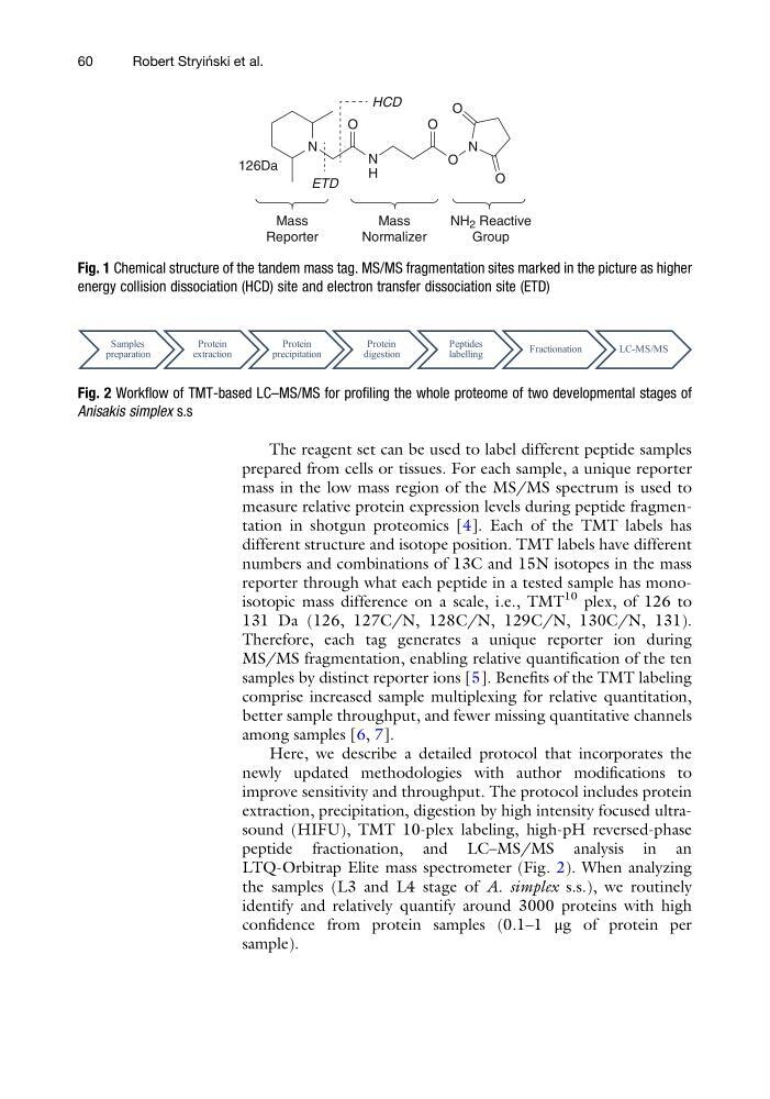
Fig. 1 Chemical structure of the tandem mass tag. MS/MS fragmentation sites marked in the picture as higher
energy collision dissociation (HCD) site and electron transfer dissociation site (ETD)
Fig. 2 Workflow of TMT-based LC–MS/MS for profiling the whole proteome of two developmental stages of
Anisakis simplex s.s
60
Robert Stryin ´ ski et al.
prepared from cells or tissues. For each sample, a unique reporter
mass in the low mass region of the MS/MS spectrum is used to
measure relative protein expression levels during peptide fragmentation in shotgun proteomics [4]. Each of the TMT labels has
different structure and isotope position. TMT labels have different
numbers and combinations of 13C and 15N isotopes in the mass
reporter through what each peptide in a tested sample has monoisotopic mass difference on a scale, i.e., TMT
10 plex, of 126 to
131 Da (126, 127C/N, 128C/N, 129C/N, 130C/N, 131).
Therefore, each tag generates a unique reporter ion during
MS/MS fragmentation, enabling relative quantification of the ten
samples by distinct reporter ions [5]. Benefits of the TMT labeling
comprise increased sample multiplexing for relative quantitation,
better sample throughput, and fewer missing quantitative channels
among samples [6, 7].
Here, we describe a detailed protocol that incorporates the
newly updated methodologies with author modifications to
improve sensitivity and throughput. The protocol includes protein
extraction, precipitation, digestion by high intensity focused ultrasound (HIFU), TMT 10-plex labeling, high-pH reversed-phase
peptide fractionation, and LC–MS/MS analysis in an
LTQ-Orbitrap Elite mass spectrometer (Fig. 2). When analyzing
the samples (L3 and L4 stage of A. simplex s.s.), we routinely
identify and relatively quantify around 3000 proteins with high
confidence from protein samples (0.1–1 μg of protein per
sample).
N
N
H
N
126Da
O
O
O
O
O
ETD
HCD
Mass
Reporter
Mass
Normalizer
NH 2 Reactive
Group
Fig. 1 Chemical structure of the tandem mass tag. MS/MS fragmentation sites marked in the picture as higher
energy collision dissociation (HCD) site and electron transfer dissociation site (ETD)
Fig. 2 Workflow of TMT-based LC–MS/MS for profiling the whole proteome of two developmental stages of
Anisakis simplex s.s
60
Robert Stryin ´ ski et al.