

6. Transfer the supernatant into 1.5-mL tubes and centrifuge
them at 15,000 Â g for 2 min using a fixed-angle centrifuge
to obtain PFP (platelet-free plasma).
7. Freeze the samples at À80
C until EV isolation (see Note 4).
3.2 EV Isolation
Plasma EV isolation follows a protocol developed in our group
based on the protocol initially established by Ramacciotti and
colleagues [8, 9, 11].
1. Add 1.2 mL of PFP per tube and mix it with 3 mL of HEPES
buffer. In total, 7.4 mL of plasma is used per run. Ultracentrifuge them at 200,000 Â g for 90 min (4
C).
2. Remove the supernatant and resuspend the pellet with 4 mL of
0.25 M KBr. Incubate on ice for 20 min (see Note 5).
3. Spin down the samples for 90 min at 4
C, 200,000 Â g.
4. Remove the supernatant and wash the pellet with 4 mL of PBS
in order to avoid plasma proteins. Centrifugate them under the
same conditions.
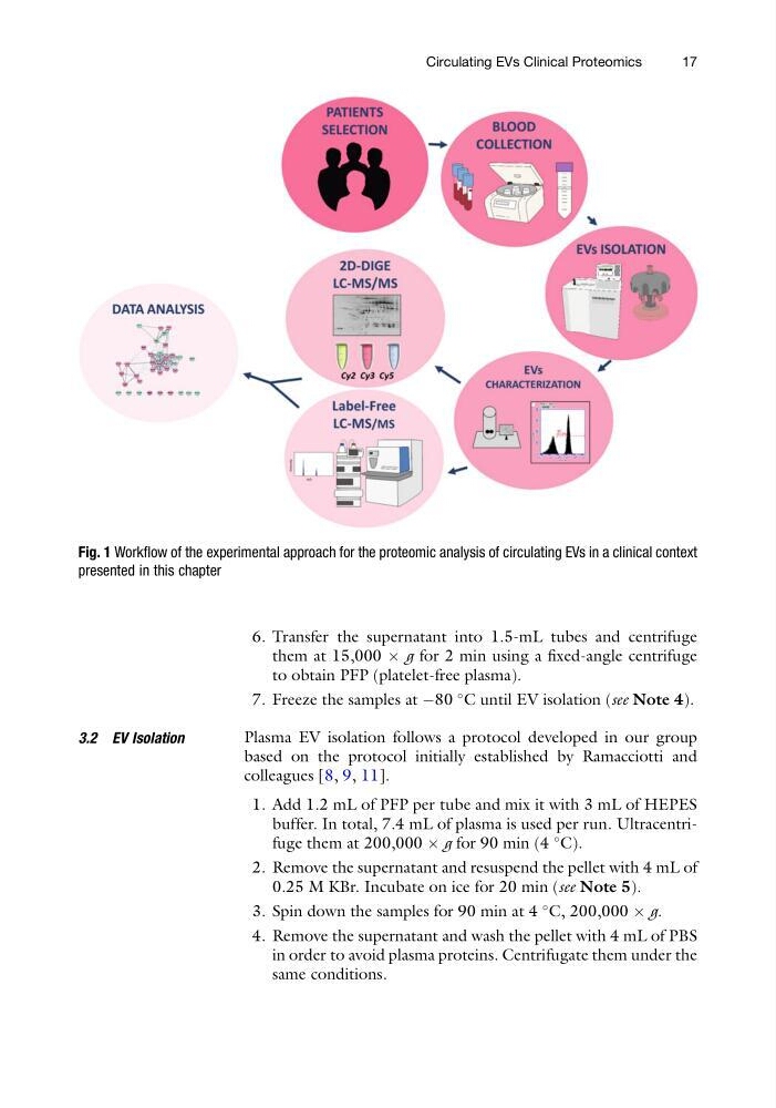
Fig. 1 Workflow of the experimental approach for the proteomic analysis of circulating EVs in a clinical context
presented in this chapter
Circulating EVs Clinical Proteomics
17
them at 15,000 Â g for 2 min using a fixed-angle centrifuge
to obtain PFP (platelet-free plasma).
7. Freeze the samples at À80
C until EV isolation (see Note 4).
3.2 EV Isolation
Plasma EV isolation follows a protocol developed in our group
based on the protocol initially established by Ramacciotti and
colleagues [8, 9, 11].
1. Add 1.2 mL of PFP per tube and mix it with 3 mL of HEPES
buffer. In total, 7.4 mL of plasma is used per run. Ultracentrifuge them at 200,000 Â g for 90 min (4
C).
2. Remove the supernatant and resuspend the pellet with 4 mL of
0.25 M KBr. Incubate on ice for 20 min (see Note 5).
3. Spin down the samples for 90 min at 4
C, 200,000 Â g.
4. Remove the supernatant and wash the pellet with 4 mL of PBS
in order to avoid plasma proteins. Centrifugate them under the
same conditions.
Fig. 1 Workflow of the experimental approach for the proteomic analysis of circulating EVs in a clinical context
presented in this chapter
Circulating EVs Clinical Proteomics
17