

Fig. 1 Workflow for MS-based phosphoproteomic analysis of cell lysate digests
(left: standard method; right: TMT-based method)
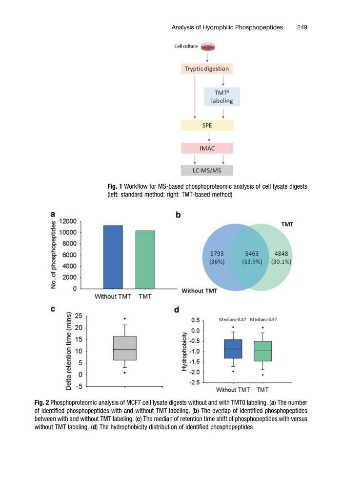
Fig. 2 Phosphoproteomic analysis of MCF7 cell lysate digests without and with TMT0 labeling. (a) The number
of identified phosphopeptides with and without TMT labeling. (b) The overlap of identified phosphopeptides
between with and without TMT labeling. (c) The median of retention time shift of phosphopeptides with versus
without TMT labeling. (d) The hydrophobicity distribution of identified phosphopeptides
Analysis of Hydrophilic Phosphopeptides
249
(left: standard method; right: TMT-based method)
Fig. 2 Phosphoproteomic analysis of MCF7 cell lysate digests without and with TMT0 labeling. (a) The number
of identified phosphopeptides with and without TMT labeling. (b) The overlap of identified phosphopeptides
between with and without TMT labeling. (c) The median of retention time shift of phosphopeptides with versus
without TMT labeling. (d) The hydrophobicity distribution of identified phosphopeptides
Analysis of Hydrophilic Phosphopeptides
249