

1. Euthanize mice in accordance with the ethical permit and
dissect out the complete colon.
2. Transfer colon to a clean silicone-covered plate and cut around
3 cm of the distal part for further mucus collection.
3. Cut the colon open lengthwise, remove any fecal pellets, and
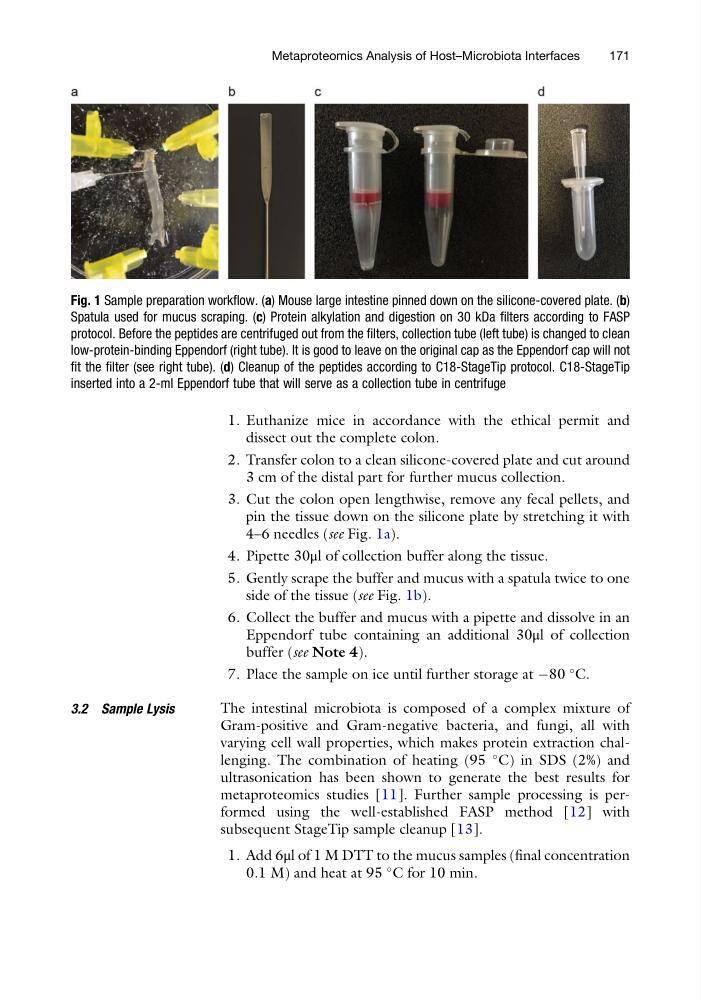
pin the tissue down on the silicone plate by stretching it with
4–6 needles (see Fig. 1a).
4. Pipette 30μl of collection buffer along the tissue.
5. Gently scrape the buffer and mucus with a spatula twice to one
side of the tissue (see Fig. 1b).
6. Collect the buffer and mucus with a pipette and dissolve in an
Eppendorf tube containing an additional 30μl of collection
buffer (see Note 4).
7. Place the sample on ice until further storage at À80
C.
3.2 Sample Lysis
The intestinal microbiota is composed of a complex mixture of
Gram-positive and Gram-negative bacteria, and fungi, all with
varying cell wall properties, which makes protein extraction challenging. The combination of heating (95
C) in SDS (2%) and
ultrasonication has been shown to generate the best results for
metaproteomics studies [11]. Further sample processing is performed using the well-established FASP method [12] with
subsequent StageTip sample cleanup [13].
1. Add 6μl of 1 M DTT to the mucus samples (final concentration
0.1 M) and heat at 95
C for 10 min.
Fig. 1 Sample preparation workflow. (a) Mouse large intestine pinned down on the silicone-covered plate. (b)
Spatula used for mucus scraping. (c) Protein alkylation and digestion on 30 kDa filters according to FASP
protocol. Before the peptides are centrifuged out from the filters, collection tube (left tube) is changed to clean
low-protein-binding Eppendorf (right tube). It is good to leave on the original cap as the Eppendorf cap will not
fit the filter (see right tube). (d) Cleanup of the peptides according to C18-StageTip protocol. C18-StageTip
inserted into a 2-ml Eppendorf tube that will serve as a collection tube in centrifuge
Metaproteomics Analysis of Host–Microbiota Interfaces
171
dissect out the complete colon.
2. Transfer colon to a clean silicone-covered plate and cut around
3 cm of the distal part for further mucus collection.
3. Cut the colon open lengthwise, remove any fecal pellets, and
pin the tissue down on the silicone plate by stretching it with
4–6 needles (see Fig. 1a).
4. Pipette 30μl of collection buffer along the tissue.
5. Gently scrape the buffer and mucus with a spatula twice to one
side of the tissue (see Fig. 1b).
6. Collect the buffer and mucus with a pipette and dissolve in an
Eppendorf tube containing an additional 30μl of collection
buffer (see Note 4).
7. Place the sample on ice until further storage at À80
C.
3.2 Sample Lysis
The intestinal microbiota is composed of a complex mixture of
Gram-positive and Gram-negative bacteria, and fungi, all with
varying cell wall properties, which makes protein extraction challenging. The combination of heating (95
C) in SDS (2%) and
ultrasonication has been shown to generate the best results for
metaproteomics studies [11]. Further sample processing is performed using the well-established FASP method [12] with
subsequent StageTip sample cleanup [13].
1. Add 6μl of 1 M DTT to the mucus samples (final concentration
0.1 M) and heat at 95
C for 10 min.
Fig. 1 Sample preparation workflow. (a) Mouse large intestine pinned down on the silicone-covered plate. (b)
Spatula used for mucus scraping. (c) Protein alkylation and digestion on 30 kDa filters according to FASP
protocol. Before the peptides are centrifuged out from the filters, collection tube (left tube) is changed to clean
low-protein-binding Eppendorf (right tube). It is good to leave on the original cap as the Eppendorf cap will not
fit the filter (see right tube). (d) Cleanup of the peptides according to C18-StageTip protocol. C18-StageTip
inserted into a 2-ml Eppendorf tube that will serve as a collection tube in centrifuge
Metaproteomics Analysis of Host–Microbiota Interfaces
171