

diluted, labeled samples to the wells and incubate at 23
C
overnight under permanent shaking on a microtiter plate
mixer.
4. Wash the beads with 3 Â 100μL wash buffer for 96-well plates
and 3 Â 60μL for 384-well plates. Add 50μL of crosslinking
solution, incubate for 10 min, and then wash the beads again.
5. Add 50μL of R-phycoerythrin (see Note 3) labeled streptavidin
to each well and incubate plates for 20 min at 23
C under
permanent shaking.
6. Finally, wash the beads with 100/60μL of wash buffer
(depending on if 96/384-well plate used) before measurement
with the Luminex instrumentation.
7. Adapt the instrumentation setting according to the bead IDs
included in the array and count at least 50 beads per bead
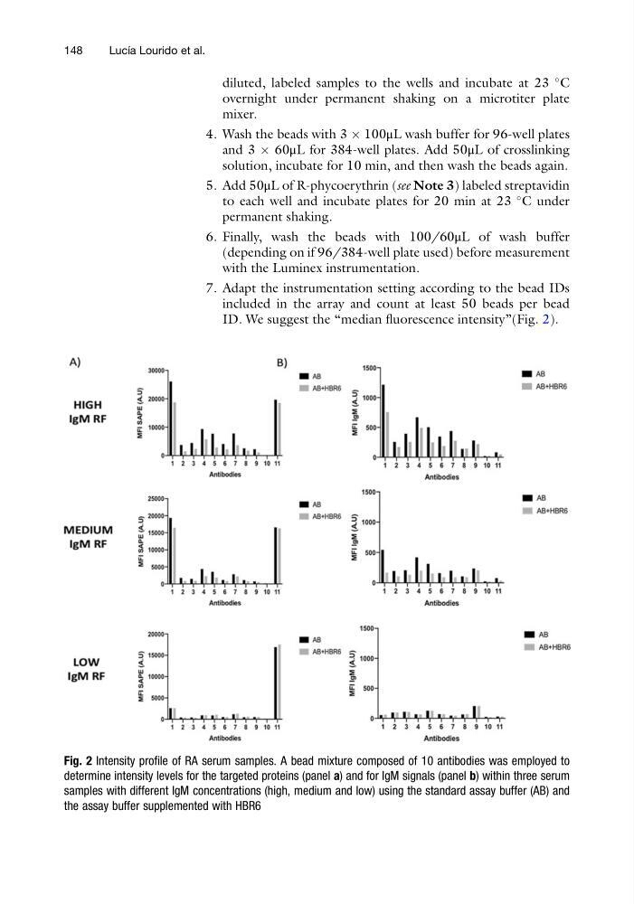
ID. We suggest the “median fluorescence intensity”(Fig. 2).
Fig. 2 Intensity profile of RA serum samples. A bead mixture composed of 10 antibodies was employed to
determine intensity levels for the targeted proteins (panel a) and for IgM signals (panel b) within three serum
samples with different IgM concentrations (high, medium and low) using the standard assay buffer (AB) and
the assay buffer supplemented with HBR6
148
Lucı ´a Lourido et al.
C
overnight under permanent shaking on a microtiter plate
mixer.
4. Wash the beads with 3 Â 100μL wash buffer for 96-well plates
and 3 Â 60μL for 384-well plates. Add 50μL of crosslinking
solution, incubate for 10 min, and then wash the beads again.
5. Add 50μL of R-phycoerythrin (see Note 3) labeled streptavidin
to each well and incubate plates for 20 min at 23
C under
permanent shaking.
6. Finally, wash the beads with 100/60μL of wash buffer
(depending on if 96/384-well plate used) before measurement
with the Luminex instrumentation.
7. Adapt the instrumentation setting according to the bead IDs
included in the array and count at least 50 beads per bead
ID. We suggest the “median fluorescence intensity”(Fig. 2).
Fig. 2 Intensity profile of RA serum samples. A bead mixture composed of 10 antibodies was employed to
determine intensity levels for the targeted proteins (panel a) and for IgM signals (panel b) within three serum
samples with different IgM concentrations (high, medium and low) using the standard assay buffer (AB) and
the assay buffer supplemented with HBR6
148
Lucı ´a Lourido et al.