

thickness, and concentrated at 60 V (constant voltage)
[115]. The run is stopped as soon as the front has penetrated
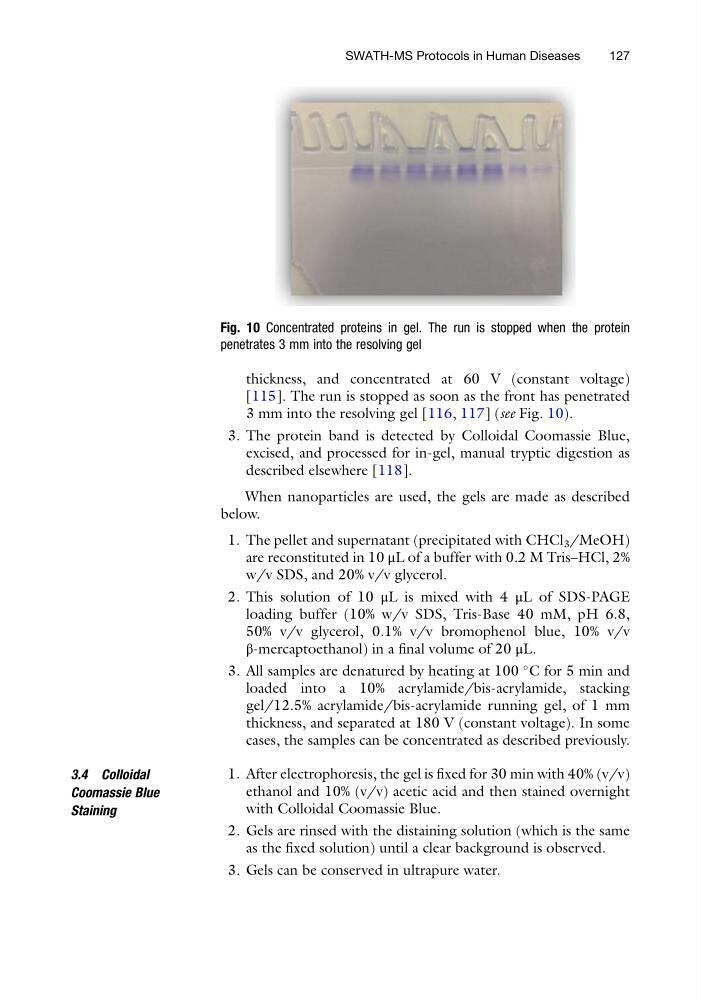
3 mm into the resolving gel [116, 117] (see Fig. 10).
3. The protein band is detected by Colloidal Coomassie Blue,
excised, and processed for in-gel, manual tryptic digestion as
described elsewhere [118].
When nanoparticles are used, the gels are made as described
below.
1. The pellet and supernatant (precipitated with CHCl 3 /MeOH)
are reconstituted in 10 μL of a buffer with 0.2 M Tris–HCl, 2%
w/v SDS, and 20% v/v glycerol.
2. This solution of 10 μL is mixed with 4 μL of SDS-PAGE
loading buffer (10% w/v SDS, Tris-Base 40 mM, pH 6.8,
50% v/v glycerol, 0.1% v/v bromophenol blue, 10% v/v
β-mercaptoethanol) in a final volume of 20 μL.
3. All samples are denatured by heating at 100
C for 5 min and
loaded into a 10% acrylamide/bis-acrylamide, stacking
gel/12.5% acrylamide/bis-acrylamide running gel, of 1 mm
thickness, and separated at 180 V (constant voltage). In some
cases, the samples can be concentrated as described previously.
3.4 Colloidal
Coomassie Blue
Staining
1. After electrophoresis, the gel is fixed for 30 min with 40% (v/v)
ethanol and 10% (v/v) acetic acid and then stained overnight
with Colloidal Coomassie Blue.
2. Gels are rinsed with the distaining solution (which is the same
as the fixed solution) until a clear background is observed.
3. Gels can be conserved in ultrapure water.
Fig. 10 Concentrated proteins in gel. The run is stopped when the protein
penetrates 3 mm into the resolving gel
SWATH-MS Protocols in Human Diseases
127
[115]. The run is stopped as soon as the front has penetrated
3 mm into the resolving gel [116, 117] (see Fig. 10).
3. The protein band is detected by Colloidal Coomassie Blue,
excised, and processed for in-gel, manual tryptic digestion as
described elsewhere [118].
When nanoparticles are used, the gels are made as described
below.
1. The pellet and supernatant (precipitated with CHCl 3 /MeOH)
are reconstituted in 10 μL of a buffer with 0.2 M Tris–HCl, 2%
w/v SDS, and 20% v/v glycerol.
2. This solution of 10 μL is mixed with 4 μL of SDS-PAGE
loading buffer (10% w/v SDS, Tris-Base 40 mM, pH 6.8,
50% v/v glycerol, 0.1% v/v bromophenol blue, 10% v/v
β-mercaptoethanol) in a final volume of 20 μL.
3. All samples are denatured by heating at 100
C for 5 min and
loaded into a 10% acrylamide/bis-acrylamide, stacking
gel/12.5% acrylamide/bis-acrylamide running gel, of 1 mm
thickness, and separated at 180 V (constant voltage). In some
cases, the samples can be concentrated as described previously.
3.4 Colloidal
Coomassie Blue
Staining
1. After electrophoresis, the gel is fixed for 30 min with 40% (v/v)
ethanol and 10% (v/v) acetic acid and then stained overnight
with Colloidal Coomassie Blue.
2. Gels are rinsed with the distaining solution (which is the same
as the fixed solution) until a clear background is observed.
3. Gels can be conserved in ultrapure water.
Fig. 10 Concentrated proteins in gel. The run is stopped when the protein
penetrates 3 mm into the resolving gel
SWATH-MS Protocols in Human Diseases
127