

(b) Four times the sample volume of cold acetone is added to
the sample for the precipitation of proteins.
(c) The tube is vortexed and incubated for 60 min at À20
C
(see Note 6).
(d) The proteins are pelleted by centrifuging for 10 min at
13,000 Â g. The acetone is removed, leaving the protein
pellet to air-dry.
(e) Then, the protein fraction is submitted to an in-gel concentration and digestion (see next section).
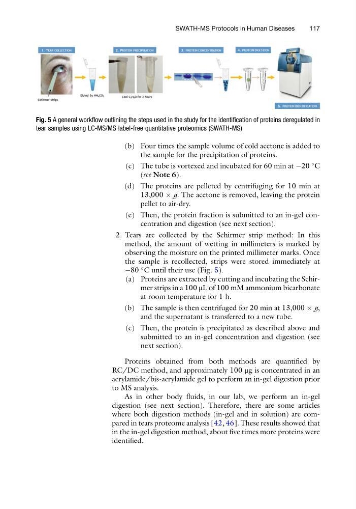
2. Tears are collected by the Schirmer strip method: In this
method, the amount of wetting in millimeters is marked by
observing the moisture on the printed millimeter marks. Once
the sample is recollected, strips were stored immediately at
À80
C until their use (Fig. 5).
(a) Proteins are extracted by cutting and incubating the Schirmer strips in a 100 μL of 100 mM ammonium bicarbonate
at room temperature for 1 h.
(b) The sample is then centrifuged for 20 min at 13,000 Â g,
and the supernatant is transferred to a new tube.
(c) Then, the protein is precipitated as described above and
submitted to an in-gel concentration and digestion (see
next section).
Proteins obtained from both methods are quantified by
RC/DC method, and approximately 100 μg is concentrated in an
acrylamide/bis-acrylamide gel to perform an in-gel digestion prior
to MS analysis.
As in other body fluids, in our lab, we perform an in-gel
digestion (see next section). Therefore, there are some articles
where both digestion methods (in-gel and in solution) are compared in tears proteome analysis [42, 46]. These results showed that
in the in-gel digestion method, about five times more proteins were
identified.
Fig. 5 A general workflow outlining the steps used in the study for the identification of proteins deregulated in
tear samples using LC-MS/MS label-free quantitative proteomics (SWATH-MS)
SWATH-MS Protocols in Human Diseases
117
the sample for the precipitation of proteins.
(c) The tube is vortexed and incubated for 60 min at À20
C
(see Note 6).
(d) The proteins are pelleted by centrifuging for 10 min at
13,000 Â g. The acetone is removed, leaving the protein
pellet to air-dry.
(e) Then, the protein fraction is submitted to an in-gel concentration and digestion (see next section).
2. Tears are collected by the Schirmer strip method: In this
method, the amount of wetting in millimeters is marked by
observing the moisture on the printed millimeter marks. Once
the sample is recollected, strips were stored immediately at
À80
C until their use (Fig. 5).
(a) Proteins are extracted by cutting and incubating the Schirmer strips in a 100 μL of 100 mM ammonium bicarbonate
at room temperature for 1 h.
(b) The sample is then centrifuged for 20 min at 13,000 Â g,
and the supernatant is transferred to a new tube.
(c) Then, the protein is precipitated as described above and
submitted to an in-gel concentration and digestion (see
next section).
Proteins obtained from both methods are quantified by
RC/DC method, and approximately 100 μg is concentrated in an
acrylamide/bis-acrylamide gel to perform an in-gel digestion prior
to MS analysis.
As in other body fluids, in our lab, we perform an in-gel
digestion (see next section). Therefore, there are some articles
where both digestion methods (in-gel and in solution) are compared in tears proteome analysis [42, 46]. These results showed that
in the in-gel digestion method, about five times more proteins were
identified.
Fig. 5 A general workflow outlining the steps used in the study for the identification of proteins deregulated in
tear samples using LC-MS/MS label-free quantitative proteomics (SWATH-MS)
SWATH-MS Protocols in Human Diseases
117