

3.1.5 In Situ Hybridization (ISH)
ISH is the only method able to provide insight into both miRNA level and localization in single cells. ISH can be divided into chromogenic enzyme-based in situ
hybridization and fluorescent in situ hybridization (FISH). miRNA ISH is technically difficult due to miRNA features such as small size, sequence similarity and low
expression levels. The standard ISH protocol has been variously modified to
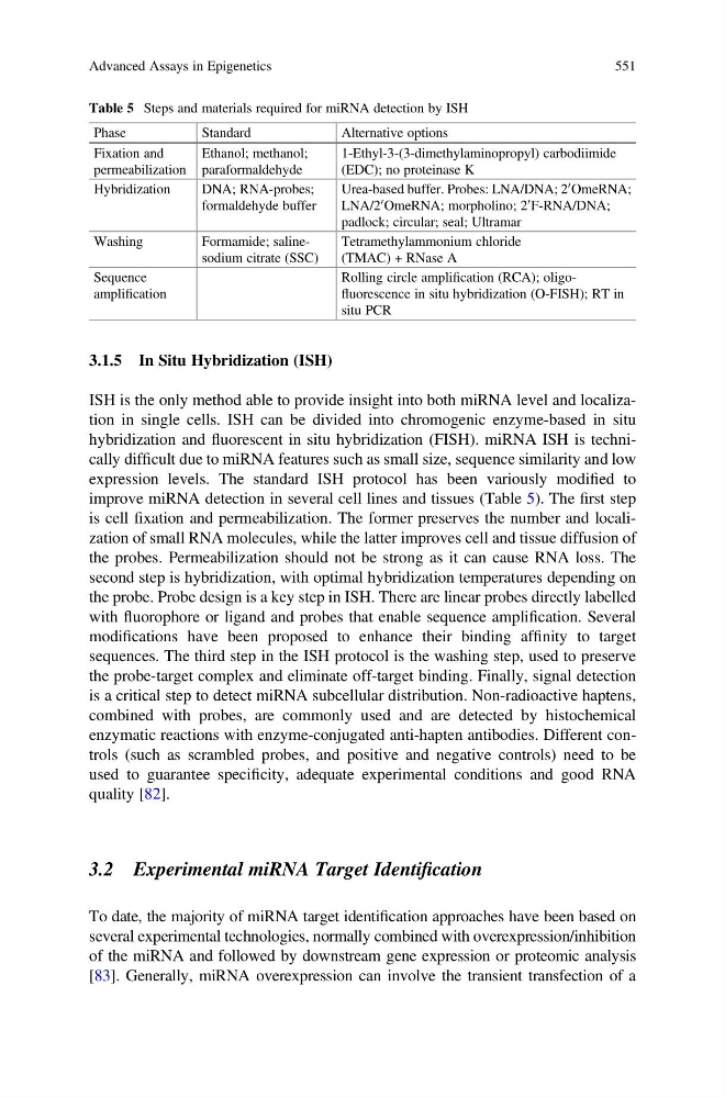
improve miRNA detection in several cell lines and tissues (Table 5). The first step
is cell fixation and permeabilization. The former preserves the number and localization of small RNA molecules, while the latter improves cell and tissue diffusion of
the probes. Permeabilization should not be strong as it can cause RNA loss. The
second step is hybridization, with optimal hybridization temperatures depending on
the probe. Probe design is a key step in ISH. There are linear probes directly labelled
with fluorophore or ligand and probes that enable sequence amplification. Several
modifications have been proposed to enhance their binding affinity to target
sequences. The third step in the ISH protocol is the washing step, used to preserve
the probe-target complex and eliminate off-target binding. Finally, signal detection
is a critical step to detect miRNA subcellular distribution. Non-radioactive haptens,
combined with probes, are commonly used and are detected by histochemical
enzymatic reactions with enzyme-conjugated anti-hapten antibodies. Different controls (such as scrambled probes, and positive and negative controls) need to be
used to guarantee specificity, adequate experimental conditions and good RNA
quality [82].
3.2 Experimental miRNA Target Identification
To date, the majority of miRNA target identification approaches have been based on
several experimental technologies, normally combined with overexpression/inhibition
of the miRNA and followed by downstream gene expression or proteomic analysis
[83]. Generally, miRNA overexpression can involve the transient transfection of a
Table 5 Steps and materials required for miRNA detection by ISH
Phase
Standard
Alternative options
Fixation and
permeabilization
Ethanol; methanol;
paraformaldehyde
1-Ethyl-3-(3-dimethylaminopropyl) carbodiimide
(EDC); no proteinase K
Hybridization
DNA; RNA-probes;
formaldehyde buffer
Urea-based buffer. Probes: LNA/DNA; 2
0 OmeRNA;
LNA/2
0 OmeRNA; morpholino; 2
0 F-RNA/DNA;
padlock; circular; seal; Ultramar
Washing
Formamide; salinesodium citrate (SSC)
Tetramethylammonium chloride
(TMAC) + RNase A
Sequence
amplification
Rolling circle amplification (RCA); oligofluorescence in situ hybridization (O-FISH); RT in
situ PCR
Advanced Assays in Epigenetics
551
ISH is the only method able to provide insight into both miRNA level and localization in single cells. ISH can be divided into chromogenic enzyme-based in situ
hybridization and fluorescent in situ hybridization (FISH). miRNA ISH is technically difficult due to miRNA features such as small size, sequence similarity and low
expression levels. The standard ISH protocol has been variously modified to
improve miRNA detection in several cell lines and tissues (Table 5). The first step
is cell fixation and permeabilization. The former preserves the number and localization of small RNA molecules, while the latter improves cell and tissue diffusion of
the probes. Permeabilization should not be strong as it can cause RNA loss. The
second step is hybridization, with optimal hybridization temperatures depending on
the probe. Probe design is a key step in ISH. There are linear probes directly labelled
with fluorophore or ligand and probes that enable sequence amplification. Several
modifications have been proposed to enhance their binding affinity to target
sequences. The third step in the ISH protocol is the washing step, used to preserve
the probe-target complex and eliminate off-target binding. Finally, signal detection
is a critical step to detect miRNA subcellular distribution. Non-radioactive haptens,
combined with probes, are commonly used and are detected by histochemical
enzymatic reactions with enzyme-conjugated anti-hapten antibodies. Different controls (such as scrambled probes, and positive and negative controls) need to be
used to guarantee specificity, adequate experimental conditions and good RNA
quality [82].
3.2 Experimental miRNA Target Identification
To date, the majority of miRNA target identification approaches have been based on
several experimental technologies, normally combined with overexpression/inhibition
of the miRNA and followed by downstream gene expression or proteomic analysis
[83]. Generally, miRNA overexpression can involve the transient transfection of a
Table 5 Steps and materials required for miRNA detection by ISH
Phase
Standard
Alternative options
Fixation and
permeabilization
Ethanol; methanol;
paraformaldehyde
1-Ethyl-3-(3-dimethylaminopropyl) carbodiimide
(EDC); no proteinase K
Hybridization
DNA; RNA-probes;
formaldehyde buffer
Urea-based buffer. Probes: LNA/DNA; 2
0 OmeRNA;
LNA/2
0 OmeRNA; morpholino; 2
0 F-RNA/DNA;
padlock; circular; seal; Ultramar
Washing
Formamide; salinesodium citrate (SSC)
Tetramethylammonium chloride
(TMAC) + RNase A
Sequence
amplification
Rolling circle amplification (RCA); oligofluorescence in situ hybridization (O-FISH); RT in
situ PCR
Advanced Assays in Epigenetics
551