

inhibition was improved as well as expanded to HDAC6, in agreement with previous
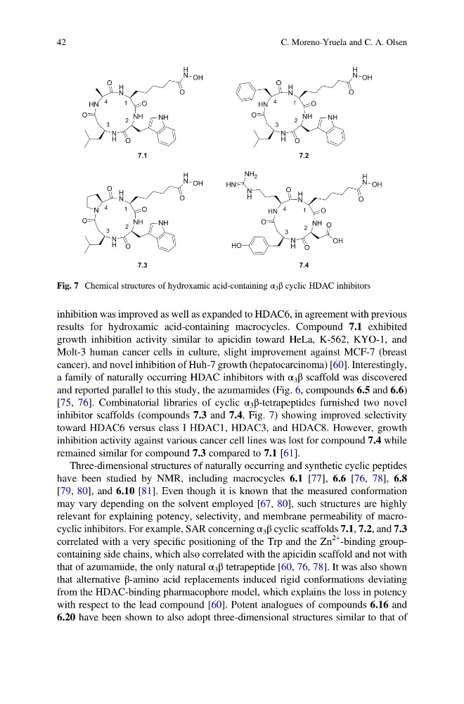
results for hydroxamic acid-containing macrocycles. Compound 7.1 exhibited
growth inhibition activity similar to apicidin toward HeLa, K-562, KYO-1, and
Molt-3 human cancer cells in culture, slight improvement against MCF-7 (breast
cancer), and novel inhibition of Huh-7 growth (hepatocarcinoma) [60]. Interestingly,
a family of naturally occurring HDAC inhibitors with α 3 β scaffold was discovered
and reported parallel to this study, the azumamides (Fig. 6, compounds 6.5 and 6.6)
[75, 76]. Combinatorial libraries of cyclic α 3 β-tetrapeptides furnished two novel
inhibitor scaffolds (compounds 7.3 and 7.4, Fig. 7) showing improved selectivity
toward HDAC6 versus class I HDAC1, HDAC3, and HDAC8. However, growth
inhibition activity against various cancer cell lines was lost for compound 7.4 while
remained similar for compound 7.3 compared to 7.1 [61].
Three-dimensional structures of naturally occurring and synthetic cyclic peptides
have been studied by NMR, including macrocycles 6.1 [77], 6.6 [76, 78], 6.8
[79, 80], and 6.10 [81]. Even though it is known that the measured conformation
may vary depending on the solvent employed [67, 80], such structures are highly
relevant for explaining potency, selectivity, and membrane permeability of macrocyclic inhibitors. For example, SAR concerning α 3 β cyclic scaffolds 7.1, 7.2, and 7.3
correlated with a very specific positioning of the Trp and the Zn
2+ -binding groupcontaining side chains, which also correlated with the apicidin scaffold and not with
that of azumamide, the only natural α 3 β tetrapeptide [60, 76, 78]. It was also shown
that alternative β-amino acid replacements induced rigid conformations deviating
from the HDAC-binding pharmacophore model, which explains the loss in potency
with respect to the lead compound [60]. Potent analogues of compounds 6.16 and
6.20 have been shown to also adopt three-dimensional structures similar to that of
HN
H
N
NH
N
H
O
O
O
NH
O
N
H
N
NH
N
H
O
O
O
NH
O
HN
H
N
NH
N
H
O
O
O
NH
O
HN
H
N
NH
N
H
O
O
O
O
OH
O
HO
7.1
7.2
7.3
7.4
O
H
N OH
O
H
N OH
O
H
N OH
O
H
N OH
N
H
HN
NH 2
1
2
3
4
1
2
3
4
1
2
3
4
1
2
3
4
Fig. 7 Chemical structures of hydroxamic acid-containing α 3 β cyclic HDAC inhibitors
42
C. Moreno-Yruela and C. A. Olsen
results for hydroxamic acid-containing macrocycles. Compound 7.1 exhibited
growth inhibition activity similar to apicidin toward HeLa, K-562, KYO-1, and
Molt-3 human cancer cells in culture, slight improvement against MCF-7 (breast
cancer), and novel inhibition of Huh-7 growth (hepatocarcinoma) [60]. Interestingly,
a family of naturally occurring HDAC inhibitors with α 3 β scaffold was discovered
and reported parallel to this study, the azumamides (Fig. 6, compounds 6.5 and 6.6)
[75, 76]. Combinatorial libraries of cyclic α 3 β-tetrapeptides furnished two novel
inhibitor scaffolds (compounds 7.3 and 7.4, Fig. 7) showing improved selectivity
toward HDAC6 versus class I HDAC1, HDAC3, and HDAC8. However, growth
inhibition activity against various cancer cell lines was lost for compound 7.4 while
remained similar for compound 7.3 compared to 7.1 [61].
Three-dimensional structures of naturally occurring and synthetic cyclic peptides
have been studied by NMR, including macrocycles 6.1 [77], 6.6 [76, 78], 6.8
[79, 80], and 6.10 [81]. Even though it is known that the measured conformation
may vary depending on the solvent employed [67, 80], such structures are highly
relevant for explaining potency, selectivity, and membrane permeability of macrocyclic inhibitors. For example, SAR concerning α 3 β cyclic scaffolds 7.1, 7.2, and 7.3
correlated with a very specific positioning of the Trp and the Zn
2+ -binding groupcontaining side chains, which also correlated with the apicidin scaffold and not with
that of azumamide, the only natural α 3 β tetrapeptide [60, 76, 78]. It was also shown
that alternative β-amino acid replacements induced rigid conformations deviating
from the HDAC-binding pharmacophore model, which explains the loss in potency
with respect to the lead compound [60]. Potent analogues of compounds 6.16 and
6.20 have been shown to also adopt three-dimensional structures similar to that of
HN
H
N
NH
N
H
O
O
O
NH
O
N
H
N
NH
N
H
O
O
O
NH
O
HN
H
N
NH
N
H
O
O
O
NH
O
HN
H
N
NH
N
H
O
O
O
O
OH
O
HO
7.1
7.2
7.3
7.4
O
H
N OH
O
H
N OH
O
H
N OH
O
H
N OH
N
H
HN
NH 2
1
2
3
4
1
2
3
4
1
2
3
4
1
2
3
4
Fig. 7 Chemical structures of hydroxamic acid-containing α 3 β cyclic HDAC inhibitors
42
C. Moreno-Yruela and C. A. Olsen