

buffer, antibody solution, and rinsing buffer. Using an antibody specific for epigenetic modifications of interest allows for determination of overall global genomic
levels. After chemiluminescence imaging, the intensity of the signal can be used to
compare the levels of epigenetic modifications between samples. Generation of
antibodies specific for different epigenetic modifications has permitted the quantification of 5mC, 5hmC, 5fC, 5caC, and N6MedA in both human and mouse tissues by
dot blot [26, 51, 52]. As is the case for ELISA-based detection, dot blots do not allow
for absolute quantification and suffer from high variability.
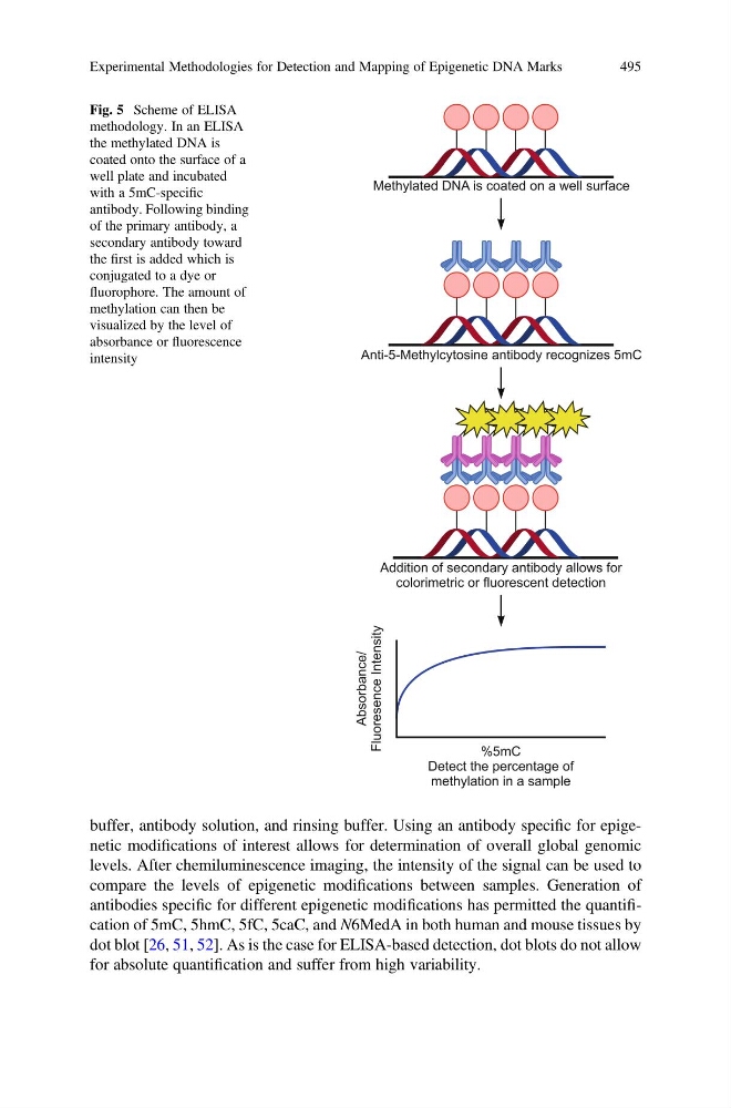
Fig. 5 Scheme of ELISA
methodology. In an ELISA
the methylated DNA is
coated onto the surface of a
well plate and incubated
with a 5mC-specific
antibody. Following binding
of the primary antibody, a
secondary antibody toward
the first is added which is
conjugated to a dye or
fluorophore. The amount of
methylation can then be
visualized by the level of
absorbance or fluorescence
intensity
Experimental Methodologies for Detection and Mapping of Epigenetic DNA Marks
495
levels. After chemiluminescence imaging, the intensity of the signal can be used to
compare the levels of epigenetic modifications between samples. Generation of
antibodies specific for different epigenetic modifications has permitted the quantification of 5mC, 5hmC, 5fC, 5caC, and N6MedA in both human and mouse tissues by
dot blot [26, 51, 52]. As is the case for ELISA-based detection, dot blots do not allow
for absolute quantification and suffer from high variability.
Fig. 5 Scheme of ELISA
methodology. In an ELISA
the methylated DNA is
coated onto the surface of a
well plate and incubated
with a 5mC-specific
antibody. Following binding
of the primary antibody, a
secondary antibody toward
the first is added which is
conjugated to a dye or
fluorophore. The amount of
methylation can then be
visualized by the level of
absorbance or fluorescence
intensity
Experimental Methodologies for Detection and Mapping of Epigenetic DNA Marks
495