

TLC was instrumental for the initial discovery of 5hmC in Purkinje cells and
cerebellum tissue [47] and for the identification of TET family of enzymes responsible for the oxidation of 5mC to 5hmC [8]. To determine the components of DNA
by TLC, the DNA was digested to nucleosides and radiolabeled with
32 P, followed
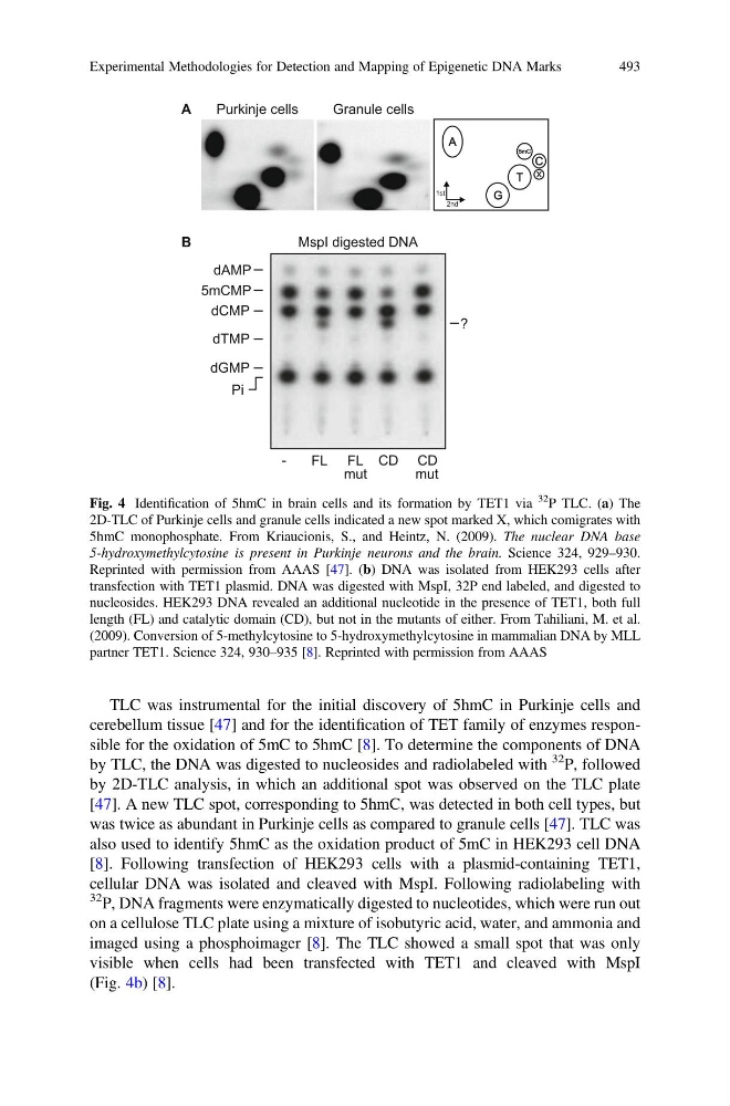
by 2D-TLC analysis, in which an additional spot was observed on the TLC plate
[47]. A new TLC spot, corresponding to 5hmC, was detected in both cell types, but
was twice as abundant in Purkinje cells as compared to granule cells [47]. TLC was
also used to identify 5hmC as the oxidation product of 5mC in HEK293 cell DNA
[8]. Following transfection of HEK293 cells with a plasmid-containing TET1,
cellular DNA was isolated and cleaved with MspI. Following radiolabeling with
32 P, DNA fragments were enzymatically digested to nucleotides, which were run out
on a cellulose TLC plate using a mixture of isobutyric acid, water, and ammonia and
imaged using a phosphoimager [8]. The TLC showed a small spot that was only
visible when cells had been transfected with TET1 and cleaved with MspI
(Fig. 4b) [8].
Fig. 4 Identification of 5hmC in brain cells and its formation by TET1 via
32
P TLC. (a) The
2D-TLC of Purkinje cells and granule cells indicated a new spot marked X, which comigrates with
5hmC monophosphate. From Kriaucionis, S., and Heintz, N. (2009). The nuclear DNA base
5-hydroxymethylcytosine is present in Purkinje neurons and the brain. Science 324, 929–930.
Reprinted with permission from AAAS [47]. (b) DNA was isolated from HEK293 cells after
transfection with TET1 plasmid. DNA was digested with MspI, 32P end labeled, and digested to
nucleosides. HEK293 DNA revealed an additional nucleotide in the presence of TET1, both full
length (FL) and catalytic domain (CD), but not in the mutants of either. From Tahiliani, M. et al.
(2009). Conversion of 5-methylcytosine to 5-hydroxymethylcytosine in mammalian DNA by MLL
partner TET1. Science 324, 930–935 [8]. Reprinted with permission from AAAS
Experimental Methodologies for Detection and Mapping of Epigenetic DNA Marks
493
cerebellum tissue [47] and for the identification of TET family of enzymes responsible for the oxidation of 5mC to 5hmC [8]. To determine the components of DNA
by TLC, the DNA was digested to nucleosides and radiolabeled with
32 P, followed
by 2D-TLC analysis, in which an additional spot was observed on the TLC plate
[47]. A new TLC spot, corresponding to 5hmC, was detected in both cell types, but
was twice as abundant in Purkinje cells as compared to granule cells [47]. TLC was
also used to identify 5hmC as the oxidation product of 5mC in HEK293 cell DNA
[8]. Following transfection of HEK293 cells with a plasmid-containing TET1,
cellular DNA was isolated and cleaved with MspI. Following radiolabeling with
32 P, DNA fragments were enzymatically digested to nucleotides, which were run out
on a cellulose TLC plate using a mixture of isobutyric acid, water, and ammonia and
imaged using a phosphoimager [8]. The TLC showed a small spot that was only
visible when cells had been transfected with TET1 and cleaved with MspI
(Fig. 4b) [8].
Fig. 4 Identification of 5hmC in brain cells and its formation by TET1 via
32
P TLC. (a) The
2D-TLC of Purkinje cells and granule cells indicated a new spot marked X, which comigrates with
5hmC monophosphate. From Kriaucionis, S., and Heintz, N. (2009). The nuclear DNA base
5-hydroxymethylcytosine is present in Purkinje neurons and the brain. Science 324, 929–930.
Reprinted with permission from AAAS [47]. (b) DNA was isolated from HEK293 cells after
transfection with TET1 plasmid. DNA was digested with MspI, 32P end labeled, and digested to
nucleosides. HEK293 DNA revealed an additional nucleotide in the presence of TET1, both full
length (FL) and catalytic domain (CD), but not in the mutants of either. From Tahiliani, M. et al.
(2009). Conversion of 5-methylcytosine to 5-hydroxymethylcytosine in mammalian DNA by MLL
partner TET1. Science 324, 930–935 [8]. Reprinted with permission from AAAS
Experimental Methodologies for Detection and Mapping of Epigenetic DNA Marks
493