

end-point assays rely on a linear behavior of the inhibitor over time, which is not the
case for slow, tight-binding molecules. Thus, preincubation times and the duration of
the assay can introduce bias in the measurement, especially when compared to faston/fast-off inhibitors.
Kinetic evidence highlights a possible ambiguity in the characterization of natural
and hydroxamic acid-containing peptide inhibitors. Previous in vitro studies with
compounds inspired by the structures of 6.3, 6.10, 6.18, and 6.20 (such as those
concerning compound 6.21) thus call for reevaluation, since only end-point inhibition data was reported. This also means that the differences observed between
in vitro HDAC inhibition and cellular antiproliferase activity for some compounds
might be derived not only from differences in membrane permeability but also
resulting from differences in mechanism. It has now been shown that potencies
measured in continuous assays can differ from other in vitro experiments and that the
mechanism of inhibition is not only class-dependent but, sometimes, even isotypedependent. Thus, investigation of the mechanism of action should be taken into
account in future studies.
Elaborating on the modification of naturally occurring macrocyclic HDAC inhibitors, further studies involved modification of the tetrapeptide scaffold. Yoshida’s
group carried out thorough structure-activity relationship (SAR) studies of the side
chain configuration of trapoxin and cyl derivatives, which interestingly revealed that,
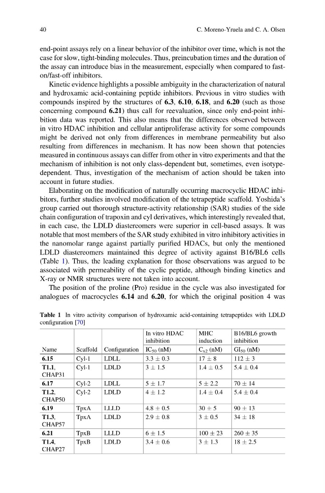
in each case, the LDLD diastereomers were superior in cell-based assays. It was
notable that most members of the SAR study exhibited in vitro inhibitory activities in
the nanomolar range against partially purified HDACs, but only the mentioned
LDLD diastereomers maintained this degree of activity against B16/BL6 cells
(Table 1). Thus, the leading explanation for those observations was argued to be
associated with permeability of the cyclic peptide, although binding kinetics and
X-ray or NMR structures were not taken into account.
The position of the proline (Pro) residue in the cycle was also investigated for
analogues of macrocycles 6.14 and 6.20, for which the original position 4 was
Table 1 In vitro activity comparison of hydroxamic acid-containing tetrapeptides with LDLD
configuration [70]
Name
Scaffold Configuration
In vitro HDAC
inhibition
MHC
induction
B16/BL6 growth
inhibition
IC 50 (nM)
C x2 (nM)
GI 50 (nM)
6.15
Cyl-1
LDLL
3.3 Æ 0.3
17 Æ 8
112 Æ 3
T1.1,
CHAP31
Cyl-1
LDLD
3 Æ 1.5
1.4 Æ 0.5
5.4 Æ 0.4
6.17
Cyl-2
LDLL
5 Æ 1.7
5 Æ 2.2
70 Æ 14
T1.2,
CHAP50
Cyl-2
LDLD
4 Æ 1.2
1.4 Æ 0.4
5.4 Æ 0.4
6.19
TpxA
LLLD
4.8 Æ 0.5
30 Æ 5
9 0Æ 13
T1.3,
CHAP57
TpxA
LDLD
2.9 Æ 0.8
3 Æ 0.5
34 Æ 18
6.21
TpxB
LLLD
6 Æ 1.5
100 Æ 23
260 Æ 35
T1.4,
CHAP27
TpxB
LDLD
3.4 Æ 0.6
3 Æ 1.3
18 Æ 2.5
40
C. Moreno-Yruela and C. A. Olsen
case for slow, tight-binding molecules. Thus, preincubation times and the duration of
the assay can introduce bias in the measurement, especially when compared to faston/fast-off inhibitors.
Kinetic evidence highlights a possible ambiguity in the characterization of natural
and hydroxamic acid-containing peptide inhibitors. Previous in vitro studies with
compounds inspired by the structures of 6.3, 6.10, 6.18, and 6.20 (such as those
concerning compound 6.21) thus call for reevaluation, since only end-point inhibition data was reported. This also means that the differences observed between
in vitro HDAC inhibition and cellular antiproliferase activity for some compounds
might be derived not only from differences in membrane permeability but also
resulting from differences in mechanism. It has now been shown that potencies
measured in continuous assays can differ from other in vitro experiments and that the
mechanism of inhibition is not only class-dependent but, sometimes, even isotypedependent. Thus, investigation of the mechanism of action should be taken into
account in future studies.
Elaborating on the modification of naturally occurring macrocyclic HDAC inhibitors, further studies involved modification of the tetrapeptide scaffold. Yoshida’s
group carried out thorough structure-activity relationship (SAR) studies of the side
chain configuration of trapoxin and cyl derivatives, which interestingly revealed that,
in each case, the LDLD diastereomers were superior in cell-based assays. It was
notable that most members of the SAR study exhibited in vitro inhibitory activities in
the nanomolar range against partially purified HDACs, but only the mentioned
LDLD diastereomers maintained this degree of activity against B16/BL6 cells
(Table 1). Thus, the leading explanation for those observations was argued to be
associated with permeability of the cyclic peptide, although binding kinetics and
X-ray or NMR structures were not taken into account.
The position of the proline (Pro) residue in the cycle was also investigated for
analogues of macrocycles 6.14 and 6.20, for which the original position 4 was
Table 1 In vitro activity comparison of hydroxamic acid-containing tetrapeptides with LDLD
configuration [70]
Name
Scaffold Configuration
In vitro HDAC
inhibition
MHC
induction
B16/BL6 growth
inhibition
IC 50 (nM)
C x2 (nM)
GI 50 (nM)
6.15
Cyl-1
LDLL
3.3 Æ 0.3
17 Æ 8
112 Æ 3
T1.1,
CHAP31
Cyl-1
LDLD
3 Æ 1.5
1.4 Æ 0.5
5.4 Æ 0.4
6.17
Cyl-2
LDLL
5 Æ 1.7
5 Æ 2.2
70 Æ 14
T1.2,
CHAP50
Cyl-2
LDLD
4 Æ 1.2
1.4 Æ 0.4
5.4 Æ 0.4
6.19
TpxA
LLLD
4.8 Æ 0.5
30 Æ 5
9 0Æ 13
T1.3,
CHAP57
TpxA
LDLD
2.9 Æ 0.8
3 Æ 0.5
34 Æ 18
6.21
TpxB
LLLD
6 Æ 1.5
100 Æ 23
260 Æ 35
T1.4,
CHAP27
TpxB
LDLD
3.4 Æ 0.6
3 Æ 1.3
18 Æ 2.5
40
C. Moreno-Yruela and C. A. Olsen