

domain-containing lysine methyltransferases and non-SET domain lysine methyltransferases. PRMTs and PKMTs share a common mechanism of action, in which
S-adenosyl-L-methionine (SAM) acts as cofactor, and hence the methyl donor, and
S-adenosylhomocysteine (SAH) is formed as a by-product [99]. Methylation occurs
on a nitrogen atom of either an arginine or lysine residue in the histone tails. This
type of modification does not alter the charge of the residue, but it affects the
basicity, hydrophobicity, and the size of the amino acid side chain which in turn
has an impact on the proteins that recognize such modifications. Indeed, despite the
subtle changes, many proteins are able to distinguish and bind specific methylation
states.
So far, more than 20 PKMTs and 11 PRMTs have been identified [100]. Nevertheless, enzymatic activity could not be demonstrated for all members [101–
103]. Over the years, different names and classifications have been attributed to
PKMTs, for instance, they have been renamed as lysine methyltransferases (KMTs)
and subdivided into eight classes [104]. Many of the methyltransferases have been
linked to diverse cellular processes and different types of cancers, and, therefore, the
development of selective inhibitors is desirable [105]. Here, we illustrate some
computational studies to highlight the utility of ligand- and structure-based methods
in the discovery of new inhibitors of histone methyltransferases.
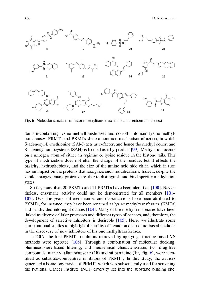
In 2007, the first PRMT1 inhibitors retrieved by applying structure-based VS
methods were reported [106]. Through a combination of molecular docking,
pharmacophore-based filtering, and biochemical characterization, two drug-like
compounds, namely, allantodapsone (18) and stilbamidine (19, Fig. 6), were identified as substrate-competitive inhibitors of PRMT1. In this study, the authors
generated a homology model of PRMT1 which was subsequently used for screening
the National Cancer Institute (NCI) diversity set into the substrate binding site.
18
NH 2
S
O
O
N
H
N
O
O
O
N
H
O
NH
NH 2
NH
N
H 2
S
O
O
NH
N
H 2
N
H
N
N
H 2
S
O
O
NH
O
N
H 2
O
N
H
O
NH 2
OH
N
H
O
N
N
H
O
N
N
NH
O
OH
N
N
N
N
NH
N
O
O
N
N
N
N
NH
N
O
O
N
O
O
S
O
N
H
NH 2
O 2 N
N
N
N
H
O
Br
19
20
21
25
24
23
22
26
27
28
29
Fig. 6 Molecular structures of histone methyltransferase inhibitors mentioned in the text
466
D. Robaa et al.
S-adenosyl-L-methionine (SAM) acts as cofactor, and hence the methyl donor, and
S-adenosylhomocysteine (SAH) is formed as a by-product [99]. Methylation occurs
on a nitrogen atom of either an arginine or lysine residue in the histone tails. This
type of modification does not alter the charge of the residue, but it affects the
basicity, hydrophobicity, and the size of the amino acid side chain which in turn
has an impact on the proteins that recognize such modifications. Indeed, despite the
subtle changes, many proteins are able to distinguish and bind specific methylation
states.
So far, more than 20 PKMTs and 11 PRMTs have been identified [100]. Nevertheless, enzymatic activity could not be demonstrated for all members [101–
103]. Over the years, different names and classifications have been attributed to
PKMTs, for instance, they have been renamed as lysine methyltransferases (KMTs)
and subdivided into eight classes [104]. Many of the methyltransferases have been
linked to diverse cellular processes and different types of cancers, and, therefore, the
development of selective inhibitors is desirable [105]. Here, we illustrate some
computational studies to highlight the utility of ligand- and structure-based methods
in the discovery of new inhibitors of histone methyltransferases.
In 2007, the first PRMT1 inhibitors retrieved by applying structure-based VS
methods were reported [106]. Through a combination of molecular docking,
pharmacophore-based filtering, and biochemical characterization, two drug-like
compounds, namely, allantodapsone (18) and stilbamidine (19, Fig. 6), were identified as substrate-competitive inhibitors of PRMT1. In this study, the authors
generated a homology model of PRMT1 which was subsequently used for screening
the National Cancer Institute (NCI) diversity set into the substrate binding site.
18
NH 2
S
O
O
N
H
N
O
O
O
N
H
O
NH
NH 2
NH
N
H 2
S
O
O
NH
N
H 2
N
H
N
N
H 2
S
O
O
NH
O
N
H 2
O
N
H
O
NH 2
OH
N
H
O
N
N
H
O
N
N
NH
O
OH
N
N
N
N
NH
N
O
O
N
N
N
N
NH
N
O
O
N
O
O
S
O
N
H
NH 2
O 2 N
N
N
N
H
O
Br
19
20
21
25
24
23
22
26
27
28
29
Fig. 6 Molecular structures of histone methyltransferase inhibitors mentioned in the text
466
D. Robaa et al.