

postulation that the inhibitor is interacting with the nicotinamide-binding pocket of
Sirt2. AGK2 inhibits Sirt2 with an IC 50 value of 3.5 μM and shows more than
tenfold selectivity over Sirt1 and Sirt3. AGK2 is a valuable chemical tool since it
also displayed the ability to block α-synuclein-mediated toxicity in a Parkinson’s
disease model, possibly by modulating tubulin acetylation regulated by Sirt2 [94].
Another approach to discover novel sirtuin inhibitors was based on fragments
identified by random screening and applying structure-based design to optimize the
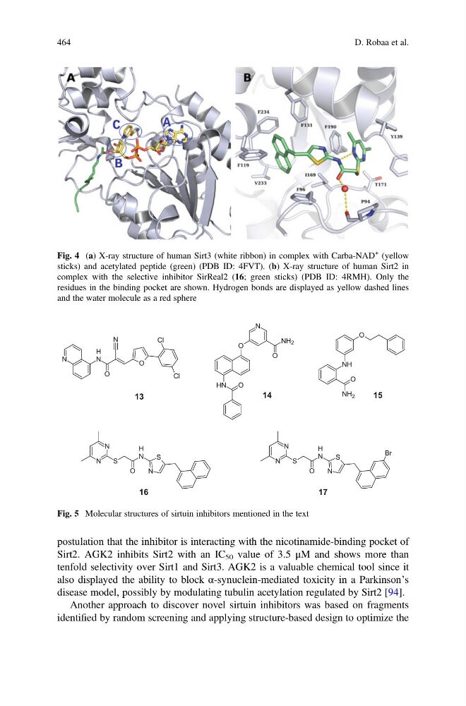
Fig. 4 (a) X-ray structure of human Sirt3 (white ribbon) in complex with Carba-NAD
+ (yellow
sticks) and acetylated peptide (green) (PDB ID: 4FVT). (b) X-ray structure of human Sirt2 in
complex with the selective inhibitor SirReal2 (16; green sticks) (PDB ID: 4RMH). Only the
residues in the binding pocket are shown. Hydrogen bonds are displayed as yellow dashed lines
and the water molecule as a red sphere
N
N
H
O
O
Cl
Cl
N
O
N
H
O
N
O
NH 2
O
N
H
N
S
S
N
N
13
14
15
O
NH
NH 2
O
16
O
N
H
N
S
S
N
N
Br
17
Fig. 5 Molecular structures of sirtuin inhibitors mentioned in the text
464
D. Robaa et al.
Sirt2. AGK2 inhibits Sirt2 with an IC 50 value of 3.5 μM and shows more than
tenfold selectivity over Sirt1 and Sirt3. AGK2 is a valuable chemical tool since it
also displayed the ability to block α-synuclein-mediated toxicity in a Parkinson’s
disease model, possibly by modulating tubulin acetylation regulated by Sirt2 [94].
Another approach to discover novel sirtuin inhibitors was based on fragments
identified by random screening and applying structure-based design to optimize the
Fig. 4 (a) X-ray structure of human Sirt3 (white ribbon) in complex with Carba-NAD
+ (yellow
sticks) and acetylated peptide (green) (PDB ID: 4FVT). (b) X-ray structure of human Sirt2 in
complex with the selective inhibitor SirReal2 (16; green sticks) (PDB ID: 4RMH). Only the
residues in the binding pocket are shown. Hydrogen bonds are displayed as yellow dashed lines
and the water molecule as a red sphere
N
N
H
O
O
Cl
Cl
N
O
N
H
O
N
O
NH 2
O
N
H
N
S
S
N
N
13
14
15
O
NH
NH 2
O
16
O
N
H
N
S
S
N
N
Br
17
Fig. 5 Molecular structures of sirtuin inhibitors mentioned in the text
464
D. Robaa et al.