

(e.g., compound 8, Fig. 1) was designed to target smHDAC8 binding pocket
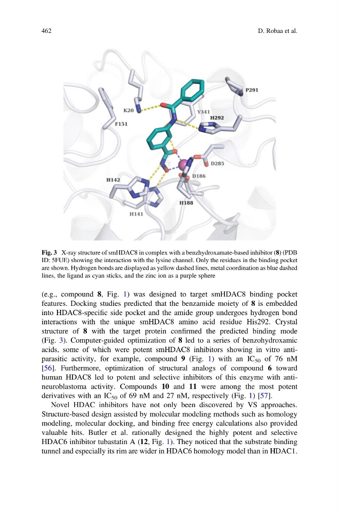
features. Docking studies predicted that the benzamide moiety of 8 is embedded
into HDAC8-specific side pocket and the amide group undergoes hydrogen bond
interactions with the unique smHDAC8 amino acid residue His292. Crystal
structure of 8 with the target protein confirmed the predicted binding mode
(Fig. 3). Computer-guided optimization of 8 led to a series of benzohydroxamic
acids, some of which were potent smHDAC8 inhibitors showing in vitro antiparasitic activity, for example, compound 9 (Fig. 1) with an IC 50 of 76 nM
[56]. Furthermore, optimization of structural analogs of compound 6 toward
human HDAC8 led to potent and selective inhibitors of this enzyme with antineuroblastoma activity. Compounds 10 and 11 were among the most potent
derivatives with an IC 50 of 69 nM and 27 nM, respectively (Fig. 1) [57].
Novel HDAC inhibitors have not only been discovered by VS approaches.
Structure-based design assisted by molecular modeling methods such as homology
modeling, molecular docking, and binding free energy calculations also provided
valuable hits. Butler et al. rationally designed the highly potent and selective
HDAC6 inhibitor tubastatin A (12, Fig. 1). They noticed that the substrate binding
tunnel and especially its rim are wider in HDAC6 homology model than in HDAC1.
Fig. 3 X-ray structure of smHDAC8 in complex with a benzhydroxamate-based inhibitor (8) (PDB
ID: 5FUE) showing the interaction with the lysine channel. Only the residues in the binding pocket
are shown. Hydrogen bonds are displayed as yellow dashed lines, metal coordination as blue dashed
lines, the ligand as cyan sticks, and the zinc ion as a purple sphere
462
D. Robaa et al.
features. Docking studies predicted that the benzamide moiety of 8 is embedded
into HDAC8-specific side pocket and the amide group undergoes hydrogen bond
interactions with the unique smHDAC8 amino acid residue His292. Crystal
structure of 8 with the target protein confirmed the predicted binding mode
(Fig. 3). Computer-guided optimization of 8 led to a series of benzohydroxamic
acids, some of which were potent smHDAC8 inhibitors showing in vitro antiparasitic activity, for example, compound 9 (Fig. 1) with an IC 50 of 76 nM
[56]. Furthermore, optimization of structural analogs of compound 6 toward
human HDAC8 led to potent and selective inhibitors of this enzyme with antineuroblastoma activity. Compounds 10 and 11 were among the most potent
derivatives with an IC 50 of 69 nM and 27 nM, respectively (Fig. 1) [57].
Novel HDAC inhibitors have not only been discovered by VS approaches.
Structure-based design assisted by molecular modeling methods such as homology
modeling, molecular docking, and binding free energy calculations also provided
valuable hits. Butler et al. rationally designed the highly potent and selective
HDAC6 inhibitor tubastatin A (12, Fig. 1). They noticed that the substrate binding
tunnel and especially its rim are wider in HDAC6 homology model than in HDAC1.
Fig. 3 X-ray structure of smHDAC8 in complex with a benzhydroxamate-based inhibitor (8) (PDB
ID: 5FUE) showing the interaction with the lysine channel. Only the residues in the binding pocket
are shown. Hydrogen bonds are displayed as yellow dashed lines, metal coordination as blue dashed
lines, the ligand as cyan sticks, and the zinc ion as a purple sphere
462
D. Robaa et al.