

The structure of human Spindlin-1 revealed a folded globular unit constituted of
a tandem of Tudor domains (Tudor 1 and Tudor 2) tightly packed against a third
Tudor domain (Tudor 3) via hydrophobic interactions, giving rise to a circular
arrangement of three Tudor domains [144]. The first and second Tudor domains
have cages made up of four aromatic residues. Spindlin-1 T1 aromatic cage binds an
H3K4me3 peptide with a K D of ~0.2 μM. Interestingly, it was found that the affinity
of the binding of Spindlin-1 to H3K4me3 is further increased by an asymmetric
methylated arginine in position 8 (H3R8me2a), which is recognized by the second
Tudor domain (K D ~0.05 μM) [145]. Furthermore, in vivo, functional experiments
suggested that Spindlin-1 activates the signaling downstream of Wnt/β-catenin from
protein arginine methyltransferase 2 (PRMT2), which creates H3R8me2a marks,
and the MLL complex, which is responsible for the generation of trimethylated
H3K4 residues. Together, this results in a specific “K4me3-R8me2a” pattern which
can be recognized by Spindlin-1 [145].
Consistent with the dual recognition of H3K4me3 and H3R8me2a, it was found
that Spindlin-1, H3K4me3, and H3R8me2a are all enriched at the promoter region
of Wnt target genes, strongly suggesting a direct role of Spindlin-1 in regulating the
expression of these genes [145]. Wnt/β-catenin signaling plays a role in a wide range
of cellular processes, including proliferation, differentiation, tissue homeostasis,
tumorigenesis, apoptosis, and cell survival. Targeting this pathway could open
new strategies for the treatment of a variety of diseases [138].
4.3.2.2 Small Molecules Targeting Tandem Tudor Domains
Targeting 53BP1
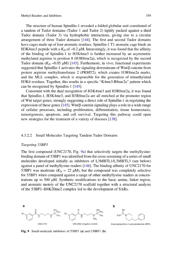
The first compound (UNC2170, Fig. 9a) that selectively targets the methyllysinebinding domain of 53BP1 was identified from the cross-screening of a series of small
molecules developed initially as inhibitors of L3MBTL1/L3MBTL3 (see below)
against a panel of methyllysine readers [146]. The binding affinity of UNC2170 for
53BP1 was moderate (K D ¼ 22 μM), but the compound was completely selective
for 53BP1 when compared against a range of other methyllysine readers at concentrations up to 500 μM. Synthetic modifications to the basic amine, linker region,
and aromatic moiety of the UNC2170 scaffold together with a structural analysis
of the 53BP1–H4K20me2 complex led to the development of SARs.
Br
N
H
O
N
H
CH 3
CH 3
CH 3
Br
N
H
O
N
CH 3
CH3
CH 3
CH 3
UNC2170
UNC2892 (negative control)
N
H 2 N
NH
4-benzylpiperidine-1-carboximidamide (BPC)
a
b
Fig. 9 Small-molecule inhibitors of 53BP1 (a) and UHRF1 (b)
Methyl-Readers and Inhibitors
359
a tandem of Tudor domains (Tudor 1 and Tudor 2) tightly packed against a third
Tudor domain (Tudor 3) via hydrophobic interactions, giving rise to a circular
arrangement of three Tudor domains [144]. The first and second Tudor domains
have cages made up of four aromatic residues. Spindlin-1 T1 aromatic cage binds an
H3K4me3 peptide with a K D of ~0.2 μM. Interestingly, it was found that the affinity
of the binding of Spindlin-1 to H3K4me3 is further increased by an asymmetric
methylated arginine in position 8 (H3R8me2a), which is recognized by the second
Tudor domain (K D ~0.05 μM) [145]. Furthermore, in vivo, functional experiments
suggested that Spindlin-1 activates the signaling downstream of Wnt/β-catenin from
protein arginine methyltransferase 2 (PRMT2), which creates H3R8me2a marks,
and the MLL complex, which is responsible for the generation of trimethylated
H3K4 residues. Together, this results in a specific “K4me3-R8me2a” pattern which
can be recognized by Spindlin-1 [145].
Consistent with the dual recognition of H3K4me3 and H3R8me2a, it was found
that Spindlin-1, H3K4me3, and H3R8me2a are all enriched at the promoter region
of Wnt target genes, strongly suggesting a direct role of Spindlin-1 in regulating the
expression of these genes [145]. Wnt/β-catenin signaling plays a role in a wide range
of cellular processes, including proliferation, differentiation, tissue homeostasis,
tumorigenesis, apoptosis, and cell survival. Targeting this pathway could open
new strategies for the treatment of a variety of diseases [138].
4.3.2.2 Small Molecules Targeting Tandem Tudor Domains
Targeting 53BP1
The first compound (UNC2170, Fig. 9a) that selectively targets the methyllysinebinding domain of 53BP1 was identified from the cross-screening of a series of small
molecules developed initially as inhibitors of L3MBTL1/L3MBTL3 (see below)
against a panel of methyllysine readers [146]. The binding affinity of UNC2170 for
53BP1 was moderate (K D ¼ 22 μM), but the compound was completely selective
for 53BP1 when compared against a range of other methyllysine readers at concentrations up to 500 μM. Synthetic modifications to the basic amine, linker region,
and aromatic moiety of the UNC2170 scaffold together with a structural analysis
of the 53BP1–H4K20me2 complex led to the development of SARs.
Br
N
H
O
N
H
CH 3
CH 3
CH 3
Br
N
H
O
N
CH 3
CH3
CH 3
CH 3
UNC2170
UNC2892 (negative control)
N
H 2 N
NH
4-benzylpiperidine-1-carboximidamide (BPC)
a
b
Fig. 9 Small-molecule inhibitors of 53BP1 (a) and UHRF1 (b)
Methyl-Readers and Inhibitors
359