

residues were also identified within these helices: three tyrosines, a phenylalanine,
an isoleucine, and two asparagines. The conserved phenylalanine was part of
what would later be termed the “WPF shelf” motif in bromodomains [20]. One of
the conserved asparagine and tyrosine residues would also later be shown to be
essential for molecular recognition of the N-ε-acetyl of acetylated lysine by
bromodomains [18].
By 1997, the family of bromodomains was expanded to include 51 bromodomains
within 37 different human bromodomain-containing proteins [21]. Using improved
structural algorithms, the domain size was also enlarged to include ~110 amino acids
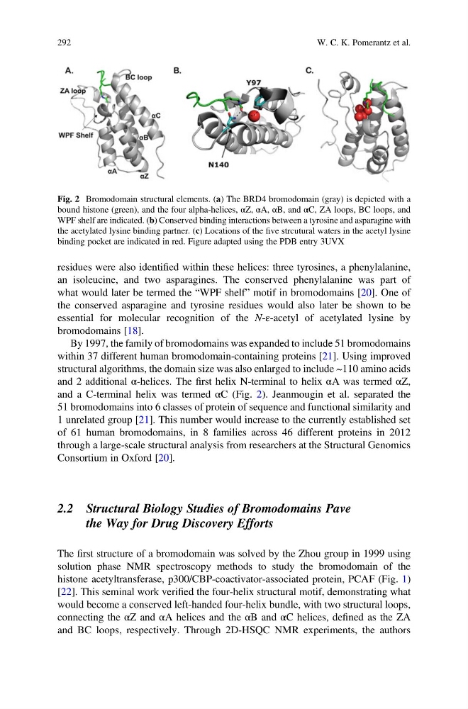
and 2 additional α-helices. The first helix N-terminal to helix αA was termed αZ,
and a C-terminal helix was termed αC (Fig. 2). Jeanmougin et al. separated the
51 bromodomains into 6 classes of protein of sequence and functional similarity and
1 unrelated group [21]. This number would increase to the currently established set
of 61 human bromodomains, in 8 families across 46 different proteins in 2012
through a large-scale structural analysis from researchers at the Structural Genomics
Consortium in Oxford [20].
2.2 Structural Biology Studies of Bromodomains Pave
the Way for Drug Discovery Efforts
The first structure of a bromodomain was solved by the Zhou group in 1999 using
solution phase NMR spectroscopy methods to study the bromodomain of the
histone acetyltransferase, p300/CBP-coactivator-associated protein, PCAF (Fig. 1)
[22]. This seminal work verified the four-helix structural motif, demonstrating what
would become a conserved left-handed four-helix bundle, with two structural loops,
connecting the αZ and αA helices and the αB and αC helices, defined as the ZA
and BC loops, respectively. Through 2D-HSQC NMR experiments, the authors
Fig. 2 Bromodomain structural elements. (a) The BRD4 bromodomain (gray) is depicted with a
bound histone (green), and the four alpha-helices, αZ, αA, αB, and αC, ZA loops, BC loops, and
WPF shelf are indicated. (b) Conserved binding interactions between a tyrosine and asparagine with
the acetylated lysine binding partner. (c) Locations of the five strcutural waters in the acetyl lysine
binding pocket are indicated in red. Figure adapted using the PDB entry 3UVX
292
W. C. K. Pomerantz et al.
an isoleucine, and two asparagines. The conserved phenylalanine was part of
what would later be termed the “WPF shelf” motif in bromodomains [20]. One of
the conserved asparagine and tyrosine residues would also later be shown to be
essential for molecular recognition of the N-ε-acetyl of acetylated lysine by
bromodomains [18].
By 1997, the family of bromodomains was expanded to include 51 bromodomains
within 37 different human bromodomain-containing proteins [21]. Using improved
structural algorithms, the domain size was also enlarged to include ~110 amino acids
and 2 additional α-helices. The first helix N-terminal to helix αA was termed αZ,
and a C-terminal helix was termed αC (Fig. 2). Jeanmougin et al. separated the
51 bromodomains into 6 classes of protein of sequence and functional similarity and
1 unrelated group [21]. This number would increase to the currently established set
of 61 human bromodomains, in 8 families across 46 different proteins in 2012
through a large-scale structural analysis from researchers at the Structural Genomics
Consortium in Oxford [20].
2.2 Structural Biology Studies of Bromodomains Pave
the Way for Drug Discovery Efforts
The first structure of a bromodomain was solved by the Zhou group in 1999 using
solution phase NMR spectroscopy methods to study the bromodomain of the
histone acetyltransferase, p300/CBP-coactivator-associated protein, PCAF (Fig. 1)
[22]. This seminal work verified the four-helix structural motif, demonstrating what
would become a conserved left-handed four-helix bundle, with two structural loops,
connecting the αZ and αA helices and the αB and αC helices, defined as the ZA
and BC loops, respectively. Through 2D-HSQC NMR experiments, the authors
Fig. 2 Bromodomain structural elements. (a) The BRD4 bromodomain (gray) is depicted with a
bound histone (green), and the four alpha-helices, αZ, αA, αB, and αC, ZA loops, BC loops, and
WPF shelf are indicated. (b) Conserved binding interactions between a tyrosine and asparagine with
the acetylated lysine binding partner. (c) Locations of the five strcutural waters in the acetyl lysine
binding pocket are indicated in red. Figure adapted using the PDB entry 3UVX
292
W. C. K. Pomerantz et al.