

Overlay of the structures of (R)-6 and (S)-6 bound in KDM5A showed differences in their binding and consequently on their potencies ((R)-6 PDB code 6BH0
and (S)-6 PDB code 6BH1) (Fig. 8d). The (S)-enantiomer engaged in extensive van
der Waals interactions between the chlorine of chlorophenyl with A411, Y409 and
Y472. The piperidine nitrogen of (R)-6 hydrogen bonds with Y472, while the
corresponding nitrogen in (S)-6 interacts with carboxylate ions of D412. The greater
binding affinity of (S)-6 could be attributed to ionic bonding with D412 if the
piperidine nitrogen is protonated. A displacement of D412 was observed between
the (R)-6 and (S)-6 structures.
A series of pyrido[3,4-d]pyrimidin-4(3H)-one inhibitors were reported by GSK
as potent KDM4 and KDM5 inhibitors as a replacement for pyridine-4-carboxylates
which suffer from poor cell permeability [66]. Several pyrazolyloxy analogues
showed good potency in biochemical assays (pIC 50 ~ 6.4) and some cellular assays
(pIC 50 ~ 5.3) against KDM4C. The best compound 7 also showed activity against
KDM5C (pIC 50 ~ 7.2) but was selective over KDM6B and EGLN3.
7 was crystallised in KDM5B (PDB:5FUN) which engaged in similar key
interactions via a monodentate coordination of pyridine nitrogen with the active
site metal and hydrogen bonding interactions of amide with K517 and N591.
Substitution at positions 5 and 6 was unfavourable due to good shape complementary at these positions. There were two conformations of Y425 which enabled
flexibility for binding to KDM5B depending on the orientation of the phenyl group.
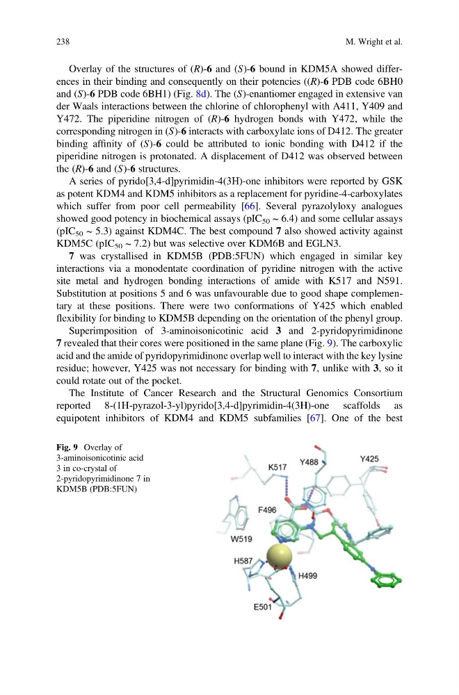
Superimposition of 3-aminoisonicotinic acid 3 and 2-pyridopyrimidinone
7 revealed that their cores were positioned in the same plane (Fig. 9). The carboxylic
acid and the amide of pyridopyrimidinone overlap well to interact with the key lysine
residue; however, Y425 was not necessary for binding with 7, unlike with 3, so it
could rotate out of the pocket.
The Institute of Cancer Research and the Structural Genomics Consortium
reported 8-(1H-pyrazol-3-yl)pyrido[3,4-d]pyrimidin-4(3H)-one scaffolds as
equipotent inhibitors of KDM4 and KDM5 subfamilies [67]. One of the best
Fig. 9 Overlay of
3-aminoisonicotinic acid
3 in co-crystal of
2-pyridopyrimidinone 7 in
KDM5B (PDB:5FUN)
238
M. Wright et al.
and (S)-6 PDB code 6BH1) (Fig. 8d). The (S)-enantiomer engaged in extensive van
der Waals interactions between the chlorine of chlorophenyl with A411, Y409 and
Y472. The piperidine nitrogen of (R)-6 hydrogen bonds with Y472, while the
corresponding nitrogen in (S)-6 interacts with carboxylate ions of D412. The greater
binding affinity of (S)-6 could be attributed to ionic bonding with D412 if the
piperidine nitrogen is protonated. A displacement of D412 was observed between
the (R)-6 and (S)-6 structures.
A series of pyrido[3,4-d]pyrimidin-4(3H)-one inhibitors were reported by GSK
as potent KDM4 and KDM5 inhibitors as a replacement for pyridine-4-carboxylates
which suffer from poor cell permeability [66]. Several pyrazolyloxy analogues
showed good potency in biochemical assays (pIC 50 ~ 6.4) and some cellular assays
(pIC 50 ~ 5.3) against KDM4C. The best compound 7 also showed activity against
KDM5C (pIC 50 ~ 7.2) but was selective over KDM6B and EGLN3.
7 was crystallised in KDM5B (PDB:5FUN) which engaged in similar key
interactions via a monodentate coordination of pyridine nitrogen with the active
site metal and hydrogen bonding interactions of amide with K517 and N591.
Substitution at positions 5 and 6 was unfavourable due to good shape complementary at these positions. There were two conformations of Y425 which enabled
flexibility for binding to KDM5B depending on the orientation of the phenyl group.
Superimposition of 3-aminoisonicotinic acid 3 and 2-pyridopyrimidinone
7 revealed that their cores were positioned in the same plane (Fig. 9). The carboxylic
acid and the amide of pyridopyrimidinone overlap well to interact with the key lysine
residue; however, Y425 was not necessary for binding with 7, unlike with 3, so it
could rotate out of the pocket.
The Institute of Cancer Research and the Structural Genomics Consortium
reported 8-(1H-pyrazol-3-yl)pyrido[3,4-d]pyrimidin-4(3H)-one scaffolds as
equipotent inhibitors of KDM4 and KDM5 subfamilies [67]. One of the best
Fig. 9 Overlay of
3-aminoisonicotinic acid
3 in co-crystal of
2-pyridopyrimidinone 7 in
KDM5B (PDB:5FUN)
238
M. Wright et al.