

to form a reactive aziridinium intermediate, able to react with the ε-NH 2 group of
lysine 79 forming a covalent bond to H3K79. This compound has not yet been
evaluated in cell-based assays. Scheufler et al. published for the first time in 2016
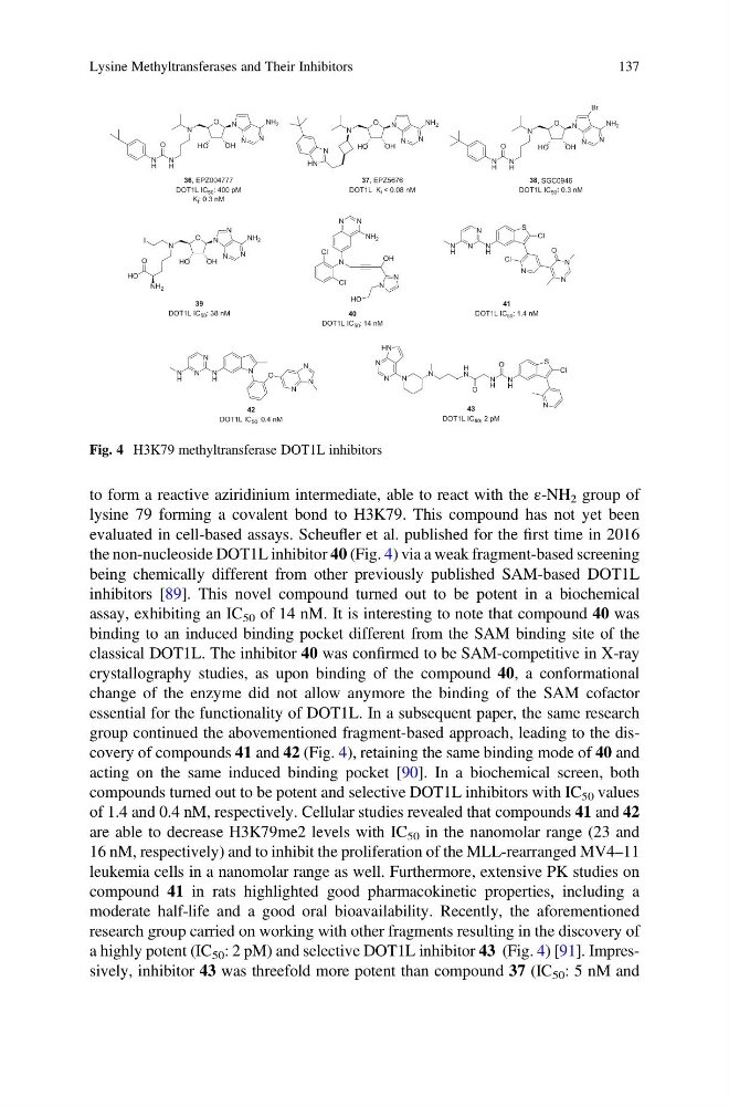
the non-nucleoside DOT1L inhibitor 40 (Fig. 4) via a weak fragment-based screening
being chemically different from other previously published SAM-based DOT1L
inhibitors [89]. This novel compound turned out to be potent in a biochemical
assay, exhibiting an IC 50 of 14 nM. It is interesting to note that compound 40 was
binding to an induced binding pocket different from the SAM binding site of the
classical DOT1L. The inhibitor 40 was confirmed to be SAM-competitive in X-ray
crystallography studies, as upon binding of the compound 40, a conformational
change of the enzyme did not allow anymore the binding of the SAM cofactor
essential for the functionality of DOT1L. In a subsequent paper, the same research
group continued the abovementioned fragment-based approach, leading to the discovery of compounds 41 and 42 (Fig. 4), retaining the same binding mode of 40 and
acting on the same induced binding pocket [90]. In a biochemical screen, both
compounds turned out to be potent and selective DOT1L inhibitors with IC 50 values
of 1.4 and 0.4 nM, respectively. Cellular studies revealed that compounds 41 and 42
are able to decrease H3K79me2 levels with IC 50 in the nanomolar range (23 and
16 nM, respectively) and to inhibit the proliferation of the MLL-rearranged MV4–11
leukemia cells in a nanomolar range as well. Furthermore, extensive PK studies on
compound 41 in rats highlighted good pharmacokinetic properties, including a
moderate half-life and a good oral bioavailability. Recently, the aforementioned
research group carried on working with other fragments resulting in the discovery of
a highly potent (IC 50 : 2 pM) and selective DOT1L inhibitor 43 (Fig. 4) [91]. Impressively, inhibitor 43 was threefold more potent than compound 37 (IC 50 : 5 nM and
N
H
N
HO
OH
N
N
N
NH 2
O
36, EPZ004777
DOT1L IC 50 : 400 pM
K i : 0.3 nM
37, EPZ5676
DOT1L Ki < 0.08 nM
38, SGC0946
DOT1L IC 50 : 0.3 nM
N
Cl
Cl
N
N
NH 2
N
N
O
N
S
N
H
N
N
N
H
Cl
Cl
N
N
H
N
N
N
H
O
N
N
N
O
H
N
N
H
O
N
H
S
N
Cl
N
N
N
N
HN
43
DOT1L IC50: 2 pM
N
H
O
N
HO
OH
N
N
N
NH 2
O
N
HN
N
HO
OH
N
N
N
NH 2
O
Br
N
H
O
N
H
N
HO
OH
N
N
N
N
NH 2
O
I
O
HO
NH 2
39
DOT1L IC 50 : 38 nM
OH
N
N
HO
42
DOT1L IC 50 : 0.4 nM
41
DOT1L IC 50 : 1.4 nM
40
DOT1L IC 50 : 14 nM
Fig. 4 H3K79 methyltransferase DOT1L inhibitors
Lysine Methyltransferases and Their Inhibitors
137
lysine 79 forming a covalent bond to H3K79. This compound has not yet been
evaluated in cell-based assays. Scheufler et al. published for the first time in 2016
the non-nucleoside DOT1L inhibitor 40 (Fig. 4) via a weak fragment-based screening
being chemically different from other previously published SAM-based DOT1L
inhibitors [89]. This novel compound turned out to be potent in a biochemical
assay, exhibiting an IC 50 of 14 nM. It is interesting to note that compound 40 was
binding to an induced binding pocket different from the SAM binding site of the
classical DOT1L. The inhibitor 40 was confirmed to be SAM-competitive in X-ray
crystallography studies, as upon binding of the compound 40, a conformational
change of the enzyme did not allow anymore the binding of the SAM cofactor
essential for the functionality of DOT1L. In a subsequent paper, the same research
group continued the abovementioned fragment-based approach, leading to the discovery of compounds 41 and 42 (Fig. 4), retaining the same binding mode of 40 and
acting on the same induced binding pocket [90]. In a biochemical screen, both
compounds turned out to be potent and selective DOT1L inhibitors with IC 50 values
of 1.4 and 0.4 nM, respectively. Cellular studies revealed that compounds 41 and 42
are able to decrease H3K79me2 levels with IC 50 in the nanomolar range (23 and
16 nM, respectively) and to inhibit the proliferation of the MLL-rearranged MV4–11
leukemia cells in a nanomolar range as well. Furthermore, extensive PK studies on
compound 41 in rats highlighted good pharmacokinetic properties, including a
moderate half-life and a good oral bioavailability. Recently, the aforementioned
research group carried on working with other fragments resulting in the discovery of
a highly potent (IC 50 : 2 pM) and selective DOT1L inhibitor 43 (Fig. 4) [91]. Impressively, inhibitor 43 was threefold more potent than compound 37 (IC 50 : 5 nM and
N
H
N
HO
OH
N
N
N
NH 2
O
36, EPZ004777
DOT1L IC 50 : 400 pM
K i : 0.3 nM
37, EPZ5676
DOT1L Ki < 0.08 nM
38, SGC0946
DOT1L IC 50 : 0.3 nM
N
Cl
Cl
N
N
NH 2
N
N
O
N
S
N
H
N
N
N
H
Cl
Cl
N
N
H
N
N
N
H
O
N
N
N
O
H
N
N
H
O
N
H
S
N
Cl
N
N
N
N
HN
43
DOT1L IC50: 2 pM
N
H
O
N
HO
OH
N
N
N
NH 2
O
N
HN
N
HO
OH
N
N
N
NH 2
O
Br
N
H
O
N
H
N
HO
OH
N
N
N
N
NH 2
O
I
O
HO
NH 2
39
DOT1L IC 50 : 38 nM
OH
N
N
HO
42
DOT1L IC 50 : 0.4 nM
41
DOT1L IC 50 : 1.4 nM
40
DOT1L IC 50 : 14 nM
Fig. 4 H3K79 methyltransferase DOT1L inhibitors
Lysine Methyltransferases and Their Inhibitors
137