

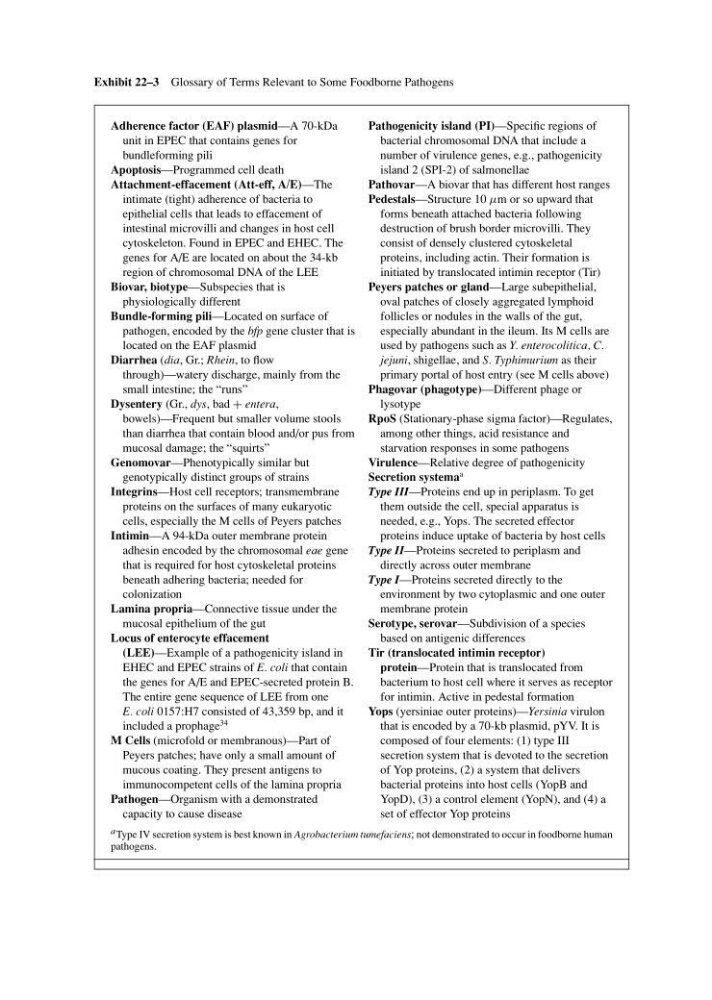
Exhibit 22–3 Glossary of Terms Relevant to Some Foodborne Pathogens
Adherence factor (EAF) plasmid—A 70-kDa
unit in EPEC that contains genes for
bundleforming pili
Apoptosis—Programmed cell death
Attachment-effacement (Att-eff, A/E)—The
intimate (tight) adherence of bacteria to
epithelial cells that leads to effacement of
intestinal microvilli and changes in host cell
cytoskeleton. Found in EPEC and EHEC. The
genes for A/E are located on about the 34-kb
region of chromosomal DNA of the LEE
Biovar, biotype—Subspecies that is
physiologically different
Bundle-forming pili—Located on surface of
pathogen, encoded by the bfp gene cluster that is
located on the EAF plasmid
Diarrhea (dia, Gr.; Rhein, to flow
through)—watery discharge, mainly from the
small intestine; the “runs”
Dysentery (Gr., dys, bad + entera,
bowels)—Frequent but smaller volume stools
than diarrhea that contain blood and/or pus from
mucosal damage; the “squirts”
Genomovar—Phenotypically similar but
genotypically distinct groups of strains
Integrins—Host cell receptors; transmembrane
proteins on the surfaces of many eukaryotic
cells, especially the M cells of Peyers patches
Intimin—A 94-kDa outer membrane protein
adhesin encoded by the chromosomal eae gene
that is required for host cytoskeletal proteins
beneath adhering bacteria; needed for
colonization
Lamina propria—Connective tissue under the
mucosal epithelium of the gut
Locus of enterocyte effacement
(LEE)—Example of a pathogenicity island in
EHEC and EPEC strains of E. coli that contain
the genes for A/E and EPEC-secreted protein B.
The entire gene sequence of LEE from one
E. coli 0157:H7 consisted of 43,359 bp, and it
included a prophage
34
M Cells (microfold or membranous)—Part of
Peyers patches; have only a small amount of
mucous coating. They present antigens to
immunocompetent cells of the lamina propria
Pathogen—Organism with a demonstrated
capacity to cause disease
Pathogenicity island (PI)—Specific regions of
bacterial chromosomal DNA that include a
number of virulence genes, e.g., pathogenicity
island 2 (SPI-2) of salmonellae
Pathovar—A biovar that has different host ranges
Pedestals—Structure 10 µm or so upward that
forms beneath attached bacteria following
destruction of brush border microvilli. They
consist of densely clustered cytoskeletal
proteins, including actin. Their formation is
initiated by translocated intimin receptor (Tir)
Peyers patches or gland—Large subepithelial,
oval patches of closely aggregated lymphoid
follicles or nodules in the walls of the gut,
especially abundant in the ileum. Its M cells are
used by pathogens such as Y. enterocolitica, C.
jejuni, shigellae, and S. Typhimurium as their
primary portal of host entry (see M cells above)
Phagovar (phagotype)—Different phage or
lysotype
RpoS (Stationary-phase sigma factor)—Regulates,
among other things, acid resistance and
starvation responses in some pathogens
Virulence—Relative degree of pathogenicity
Secretion systema
a
Type III—Proteins end up in periplasm. To get
them outside the cell, special apparatus is
needed, e.g., Yops. The secreted effector
proteins induce uptake of bacteria by host cells
Type II—Proteins secreted to periplasm and
directly across outer membrane
Type I—Proteins secreted directly to the
environment by two cytoplasmic and one outer
membrane protein
Serotype, serovar—Subdivision of a species
based on antigenic differences
Tir (translocated intimin receptor)
protein—Protein that is translocated from
bacterium to host cell where it serves as receptor
for intimin. Active in pedestal formation
Yops (yersiniae outer proteins)—Yersinia virulon
that is encoded by a 70-kb plasmid, pYV. It is
composed of four elements: (1) type III
secretion system that is devoted to the secretion
of Yop proteins, (2) a system that delivers
bacterial proteins into host cells (YopB and
YopD), (3) a control element (YopN), and (4) a
set of effector Yop proteins
a Type IV secretion system is best known in Agrobacterium tumefaciens; not demonstrated to occur in foodborne human
pathogens.