

262 Modern Food Microbiology
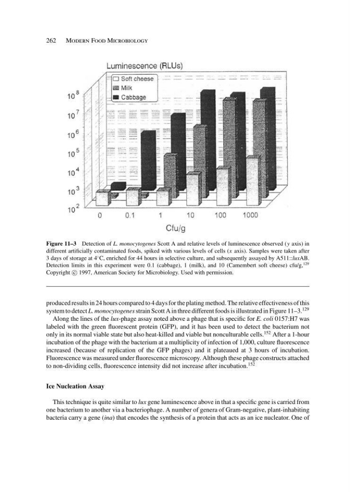
Figure 11–3 Detection of L. monocytogenes Scott A and relative levels of luminescence observed (y axis) in
different artificially contaminated foods, spiked with various levels of cells (x axis). Samples were taken after
3 days of storage at 4
◦ C, enriched for 44 hours in selective culture, and subsequently assayed by A511::luxAB.
Detection limits in this experiment were 0.1 (cabbage), 1 (milk), and 10 (Camembert soft cheese) cfu/g,
129
Copyright c
1997, American Society for Microbiology. Used with permission.
produced results in 24 hours compared to 4 days for the plating method. The relative effectiveness of this
system to detect L. monocytogenes strain Scott A in three different foods is illustrated in Figure 11–3.
129
Along the lines of the lux-phage assay noted above a phage that is specific for E. coli 0157:H7 was
labeled with the green fluorescent protein (GFP), and it has been used to detect the bacterium not
only in its normal viable state but also heat-killed and viable but nonculturable cells.
152 After a 1-hour
incubation of the phage with the bacterium at a multiplicity of infection of 1,000, culture fluorescence
increased (because of replication of the GFP phages) and it plateaued at 3 hours of incubation.
Fluorescence was measured under fluorescence microscopy. Although these phage constructs attached
to non-dividing cells, fluorescence intensity did not increase after incubation.
152
Ice Nucleation Assay
This technique is quite similar to lux gene luminescence above in that a specific gene is carried from
one bacterium to another via a bacteriophage. A number of genera of Gram-negative, plant-inhabiting
bacteria carry a gene (ina) that encodes the synthesis of a protein that acts as an ice nucleator. One of
Figure 11–3 Detection of L. monocytogenes Scott A and relative levels of luminescence observed (y axis) in
different artificially contaminated foods, spiked with various levels of cells (x axis). Samples were taken after
3 days of storage at 4
◦ C, enriched for 44 hours in selective culture, and subsequently assayed by A511::luxAB.
Detection limits in this experiment were 0.1 (cabbage), 1 (milk), and 10 (Camembert soft cheese) cfu/g,
129
Copyright c
1997, American Society for Microbiology. Used with permission.
produced results in 24 hours compared to 4 days for the plating method. The relative effectiveness of this
system to detect L. monocytogenes strain Scott A in three different foods is illustrated in Figure 11–3.
129
Along the lines of the lux-phage assay noted above a phage that is specific for E. coli 0157:H7 was
labeled with the green fluorescent protein (GFP), and it has been used to detect the bacterium not
only in its normal viable state but also heat-killed and viable but nonculturable cells.
152 After a 1-hour
incubation of the phage with the bacterium at a multiplicity of infection of 1,000, culture fluorescence
increased (because of replication of the GFP phages) and it plateaued at 3 hours of incubation.
Fluorescence was measured under fluorescence microscopy. Although these phage constructs attached
to non-dividing cells, fluorescence intensity did not increase after incubation.
152
Ice Nucleation Assay
This technique is quite similar to lux gene luminescence above in that a specific gene is carried from
one bacterium to another via a bacteriophage. A number of genera of Gram-negative, plant-inhabiting
bacteria carry a gene (ina) that encodes the synthesis of a protein that acts as an ice nucleator. One of