

10. Switch on UV light.
11. Photograph the DNA seen with an orange color bands.
5.3 Quantification
of DNA
1. Quantify the genomic DNA and amplified products using UV
spectrophotometer.
2. Read the diluted DNA sample (1:250) at 260 nm and take
distilled water as blank.
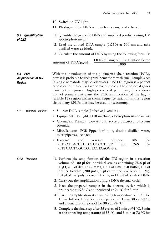
3. Calculate the amount of DNA by using the following formula:
Amount of DNA μg=μl
ð
Þ¼
OD 260 nm
ð
ÞÂ50 Â Dilution factor
1000
5.4 PCR
Amplification of ITS
Region
With the introduction of the polymerase chain reaction (PCR),
now it is probable to recognize nematodes with small sample sizes
(a single nematode may be adequate). The ITS region is a perfect
candidate for molecular taxonomic purposes. The ribosomal genes
flanking this region are highly conserved, permitting the construction of primers that assist the PCR amplification of the highly
variable ITS region within them. Sequence variation in this region
yields many RFLPs that may be used for taxonomy.
5.4.1 Materials Required
l
Source: DNA sample (Infective juveniles).
l
Equipment: UV light, PCR machine, electrophoresis apparatus.
l
Chemicals: Primers (forward and reverse), agarose, ethidium
bromide.
l
Miscellaneous: PCR Eppendorf tube, double distilled water,
micropipettes, ice pack.
l
Forward
and
reverse
primers:
18S
(50 -TTGATTACGTCCCTGCCCTTT3
0 )
and
26S
(50 -TTTCACTCGCCGTTACTAAGG-3
0 ).
5.4.2 Procedure
1. Perform the amplification of the ITS region in a reaction
volume of 100 μl for individual strains containing 75.6 μl of
H 2 O, 2 μl of dNTPs (2 mM), 10 μl of 10Â PCR buffer, 1 μl of
primer forward (200 μM), 1 μl of primer reverse (200 μM),
0.4 μl of Taq polymerase (5 U/μl), and 10 μl of purified DNA.
2. Carry out the amplification using a DNA thermal cycler.
3. Place the prepared samples in the thermal cycler, which is
pre-heated to 95
C and incubated at 94
C for 3 min.
4. Start the amplification at an annealing temperature of 55
C for
1 min, followed by an extension period for 1 min 30 s at 72
C
and a denaturation period for 30 s at 94
C.
5. Complete the final step after 35 cycles, of 1 min at 94
C, 3 min
at the annealing temperature of 55
C, and 5 min at 72
C for
Molecular Characterization
89
11. Photograph the DNA seen with an orange color bands.
5.3 Quantification
of DNA
1. Quantify the genomic DNA and amplified products using UV
spectrophotometer.
2. Read the diluted DNA sample (1:250) at 260 nm and take
distilled water as blank.
3. Calculate the amount of DNA by using the following formula:
Amount of DNA μg=μl
ð
Þ¼
OD 260 nm
ð
ÞÂ50 Â Dilution factor
1000
5.4 PCR
Amplification of ITS
Region
With the introduction of the polymerase chain reaction (PCR),
now it is probable to recognize nematodes with small sample sizes
(a single nematode may be adequate). The ITS region is a perfect
candidate for molecular taxonomic purposes. The ribosomal genes
flanking this region are highly conserved, permitting the construction of primers that assist the PCR amplification of the highly
variable ITS region within them. Sequence variation in this region
yields many RFLPs that may be used for taxonomy.
5.4.1 Materials Required
l
Source: DNA sample (Infective juveniles).
l
Equipment: UV light, PCR machine, electrophoresis apparatus.
l
Chemicals: Primers (forward and reverse), agarose, ethidium
bromide.
l
Miscellaneous: PCR Eppendorf tube, double distilled water,
micropipettes, ice pack.
l
Forward
and
reverse
primers:
18S
(50 -TTGATTACGTCCCTGCCCTTT3
0 )
and
26S
(50 -TTTCACTCGCCGTTACTAAGG-3
0 ).
5.4.2 Procedure
1. Perform the amplification of the ITS region in a reaction
volume of 100 μl for individual strains containing 75.6 μl of
H 2 O, 2 μl of dNTPs (2 mM), 10 μl of 10Â PCR buffer, 1 μl of
primer forward (200 μM), 1 μl of primer reverse (200 μM),
0.4 μl of Taq polymerase (5 U/μl), and 10 μl of purified DNA.
2. Carry out the amplification using a DNA thermal cycler.
3. Place the prepared samples in the thermal cycler, which is
pre-heated to 95
C and incubated at 94
C for 3 min.
4. Start the amplification at an annealing temperature of 55
C for
1 min, followed by an extension period for 1 min 30 s at 72
C
and a denaturation period for 30 s at 94
C.
5. Complete the final step after 35 cycles, of 1 min at 94
C, 3 min
at the annealing temperature of 55
C, and 5 min at 72
C for
Molecular Characterization
89