

46
D. Li et al.
a
b
c
d
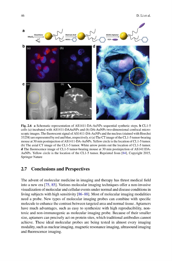
Fig. 2.6 a Schematic representation of AS1411-DA-AuNPs sequential synthetic steps. b CL1-5
cells (a) incubated with AS1411-DAAuNPs and (b) DA-AuNPs two-dimensional confocal microscopic images. The fluorescent signal of AS1411-DA-AuNPs and the nucleus (stained with Hoechst
33258) are represented by red and blue, respectively. c (a) The CT image of the CL1-5 tumor-bearing
mouse at 30 min postinjection of AS1411-DA-AuNPs. Yellow circle is the location of CL1-5 tumor.
(b) The axial CT image of the CL1-5 tumor. White arrow points out the location of CL1-5 tumor.
d The fluorescence image of CL1-5 tumor-bearing mouse at 30 min postinjection of AS1411DAAuNPs. Yellow circle is the location of the CL1-5 tumor. Reprinted from [84], Copyright 2015,
Springer Nature
2.7 Conclusions and Perspectives
The advent of molecular medicine in imaging and therapy has thrust medical field
into a new era [75, 85]. Various molecular imaging techniques offer a non-invasive
visualization of molecular and cellular events under normal and disease conditions in
living subjects with high sensitivity [86–88]. Most of molecular imaging modalities
need a probe. New types of molecular imaging probes can combine with specific
molecule to enhance the contrast between targeted area and normal tissue. Aptamers
have much advantages, such as easy to synthesize with high reproducibility, nontoxic and non-immunogenic as molecular imaging probe. Because of their smaller
size, aptamers can precisely act on protein sites, which traditional antibodies cannot
achieve. These ideal molecular probes are being tested in almost every imaging
modality, such as nuclear imaging, magnetic resonance imaging, ultrasound imaging
and fluorescence imaging.
D. Li et al.
a
b
c
d
Fig. 2.6 a Schematic representation of AS1411-DA-AuNPs sequential synthetic steps. b CL1-5
cells (a) incubated with AS1411-DAAuNPs and (b) DA-AuNPs two-dimensional confocal microscopic images. The fluorescent signal of AS1411-DA-AuNPs and the nucleus (stained with Hoechst
33258) are represented by red and blue, respectively. c (a) The CT image of the CL1-5 tumor-bearing
mouse at 30 min postinjection of AS1411-DA-AuNPs. Yellow circle is the location of CL1-5 tumor.
(b) The axial CT image of the CL1-5 tumor. White arrow points out the location of CL1-5 tumor.
d The fluorescence image of CL1-5 tumor-bearing mouse at 30 min postinjection of AS1411DAAuNPs. Yellow circle is the location of the CL1-5 tumor. Reprinted from [84], Copyright 2015,
Springer Nature
2.7 Conclusions and Perspectives
The advent of molecular medicine in imaging and therapy has thrust medical field
into a new era [75, 85]. Various molecular imaging techniques offer a non-invasive
visualization of molecular and cellular events under normal and disease conditions in
living subjects with high sensitivity [86–88]. Most of molecular imaging modalities
need a probe. New types of molecular imaging probes can combine with specific
molecule to enhance the contrast between targeted area and normal tissue. Aptamers
have much advantages, such as easy to synthesize with high reproducibility, nontoxic and non-immunogenic as molecular imaging probe. Because of their smaller
size, aptamers can precisely act on protein sites, which traditional antibodies cannot
achieve. These ideal molecular probes are being tested in almost every imaging
modality, such as nuclear imaging, magnetic resonance imaging, ultrasound imaging
and fluorescence imaging.