

44
D. Li et al.
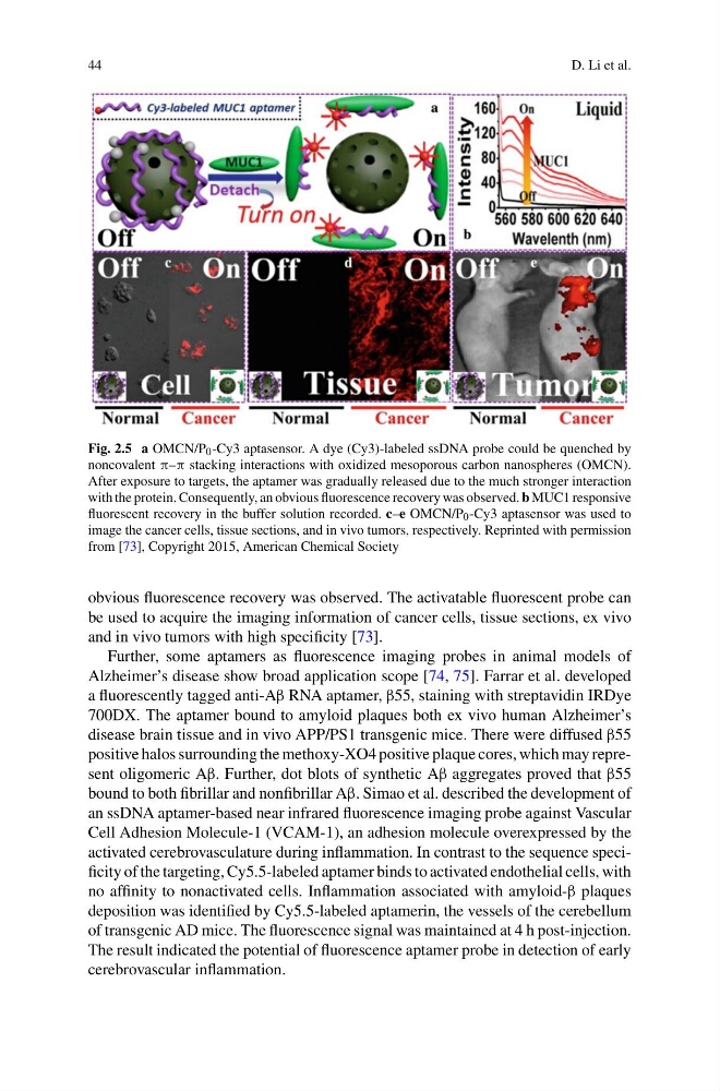
Fig. 2.5 a OMCN/P 0 -Cy3 aptasensor. A dye (Cy3)-labeled ssDNA probe could be quenched by
noncovalent π–π stacking interactions with oxidized mesoporous carbon nanospheres (OMCN).
After exposure to targets, the aptamer was gradually released due to the much stronger interaction
with the protein. Consequently, an obvious fluorescence recovery was observed. b MUC1 responsive
fluorescent recovery in the buffer solution recorded. c–e OMCN/P 0 -Cy3 aptasensor was used to
image the cancer cells, tissue sections, and in vivo tumors, respectively. Reprinted with permission
from [73], Copyright 2015, American Chemical Society
obvious fluorescence recovery was observed. The activatable fluorescent probe can
be used to acquire the imaging information of cancer cells, tissue sections, ex vivo
and in vivo tumors with high specificity [73].
Further, some aptamers as fluorescence imaging probes in animal models of
Alzheimer’s disease show broad application scope [74, 75]. Farrar et al. developed
a fluorescently tagged anti-Aβ RNA aptamer, β55, staining with streptavidin IRDye
700DX. The aptamer bound to amyloid plaques both ex vivo human Alzheimer’s
disease brain tissue and in vivo APP/PS1 transgenic mice. There were diffused β55
positive halos surrounding the methoxy-XO4 positive plaque cores, which may represent oligomeric Aβ. Further, dot blots of synthetic Aβ aggregates proved that β55
bound to both fibrillar and nonfibrillar Aβ. Simao et al. described the development of
an ssDNA aptamer-based near infrared fluorescence imaging probe against Vascular
Cell Adhesion Molecule-1 (VCAM-1), an adhesion molecule overexpressed by the
activated cerebrovasculature during inflammation. In contrast to the sequence specificity of the targeting, Cy5.5-labeled aptamer binds to activated endothelial cells, with
no affinity to nonactivated cells. Inflammation associated with amyloid-β plaques
deposition was identified by Cy5.5-labeled aptamerin, the vessels of the cerebellum
of transgenic AD mice. The fluorescence signal was maintained at 4 h post-injection.
The result indicated the potential of fluorescence aptamer probe in detection of early
cerebrovascular inflammation.
D. Li et al.
Fig. 2.5 a OMCN/P 0 -Cy3 aptasensor. A dye (Cy3)-labeled ssDNA probe could be quenched by
noncovalent π–π stacking interactions with oxidized mesoporous carbon nanospheres (OMCN).
After exposure to targets, the aptamer was gradually released due to the much stronger interaction
with the protein. Consequently, an obvious fluorescence recovery was observed. b MUC1 responsive
fluorescent recovery in the buffer solution recorded. c–e OMCN/P 0 -Cy3 aptasensor was used to
image the cancer cells, tissue sections, and in vivo tumors, respectively. Reprinted with permission
from [73], Copyright 2015, American Chemical Society
obvious fluorescence recovery was observed. The activatable fluorescent probe can
be used to acquire the imaging information of cancer cells, tissue sections, ex vivo
and in vivo tumors with high specificity [73].
Further, some aptamers as fluorescence imaging probes in animal models of
Alzheimer’s disease show broad application scope [74, 75]. Farrar et al. developed
a fluorescently tagged anti-Aβ RNA aptamer, β55, staining with streptavidin IRDye
700DX. The aptamer bound to amyloid plaques both ex vivo human Alzheimer’s
disease brain tissue and in vivo APP/PS1 transgenic mice. There were diffused β55
positive halos surrounding the methoxy-XO4 positive plaque cores, which may represent oligomeric Aβ. Further, dot blots of synthetic Aβ aggregates proved that β55
bound to both fibrillar and nonfibrillar Aβ. Simao et al. described the development of
an ssDNA aptamer-based near infrared fluorescence imaging probe against Vascular
Cell Adhesion Molecule-1 (VCAM-1), an adhesion molecule overexpressed by the
activated cerebrovasculature during inflammation. In contrast to the sequence specificity of the targeting, Cy5.5-labeled aptamer binds to activated endothelial cells, with
no affinity to nonactivated cells. Inflammation associated with amyloid-β plaques
deposition was identified by Cy5.5-labeled aptamerin, the vessels of the cerebellum
of transgenic AD mice. The fluorescence signal was maintained at 4 h post-injection.
The result indicated the potential of fluorescence aptamer probe in detection of early
cerebrovascular inflammation.