

334
Y. Cheng and H. Jin
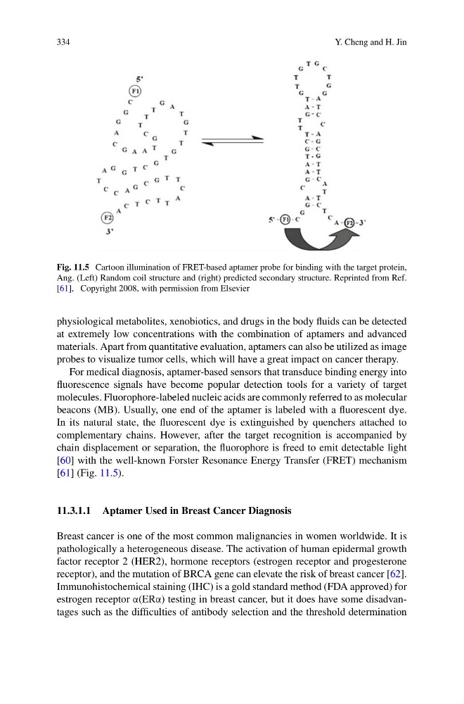
Fig. 11.5 Cartoon illumination of FRET-based aptamer probe for binding with the target protein,
Ang. (Left) Random coil structure and (right) predicted secondary structure. Reprinted from Ref.
[61], Copyright 2008, with permission from Elsevier
physiological metabolites, xenobiotics, and drugs in the body fluids can be detected
at extremely low concentrations with the combination of aptamers and advanced
materials. Apart from quantitative evaluation, aptamers can also be utilized as image
probes to visualize tumor cells, which will have a great impact on cancer therapy.
For medical diagnosis, aptamer-based sensors that transduce binding energy into
fluorescence signals have become popular detection tools for a variety of target
molecules. Fluorophore-labeled nucleic acids are commonly referred to as molecular
beacons (MB). Usually, one end of the aptamer is labeled with a fluorescent dye.
In its natural state, the fluorescent dye is extinguished by quenchers attached to
complementary chains. However, after the target recognition is accompanied by
chain displacement or separation, the fluorophore is freed to emit detectable light
[60] with the well-known Forster Resonance Energy Transfer (FRET) mechanism
[61] (Fig. 11.5).
11.3.1.1 Aptamer Used in Breast Cancer Diagnosis
Breast cancer is one of the most common malignancies in women worldwide. It is
pathologically a heterogeneous disease. The activation of human epidermal growth
factor receptor 2 (HER2), hormone receptors (estrogen receptor and progesterone
receptor), and the mutation of BRCA gene can elevate the risk of breast cancer [62].
Immunohistochemical staining (IHC) is a gold standard method (FDA approved) for
estrogen receptor α(ERα) testing in breast cancer, but it does have some disadvantages such as the difficulties of antibody selection and the threshold determination
Y. Cheng and H. Jin
Fig. 11.5 Cartoon illumination of FRET-based aptamer probe for binding with the target protein,
Ang. (Left) Random coil structure and (right) predicted secondary structure. Reprinted from Ref.
[61], Copyright 2008, with permission from Elsevier
physiological metabolites, xenobiotics, and drugs in the body fluids can be detected
at extremely low concentrations with the combination of aptamers and advanced
materials. Apart from quantitative evaluation, aptamers can also be utilized as image
probes to visualize tumor cells, which will have a great impact on cancer therapy.
For medical diagnosis, aptamer-based sensors that transduce binding energy into
fluorescence signals have become popular detection tools for a variety of target
molecules. Fluorophore-labeled nucleic acids are commonly referred to as molecular
beacons (MB). Usually, one end of the aptamer is labeled with a fluorescent dye.
In its natural state, the fluorescent dye is extinguished by quenchers attached to
complementary chains. However, after the target recognition is accompanied by
chain displacement or separation, the fluorophore is freed to emit detectable light
[60] with the well-known Forster Resonance Energy Transfer (FRET) mechanism
[61] (Fig. 11.5).
11.3.1.1 Aptamer Used in Breast Cancer Diagnosis
Breast cancer is one of the most common malignancies in women worldwide. It is
pathologically a heterogeneous disease. The activation of human epidermal growth
factor receptor 2 (HER2), hormone receptors (estrogen receptor and progesterone
receptor), and the mutation of BRCA gene can elevate the risk of breast cancer [62].
Immunohistochemical staining (IHC) is a gold standard method (FDA approved) for
estrogen receptor α(ERα) testing in breast cancer, but it does have some disadvantages such as the difficulties of antibody selection and the threshold determination