

11 Hormone Aptamers in Endocrine-Related Diseases
331
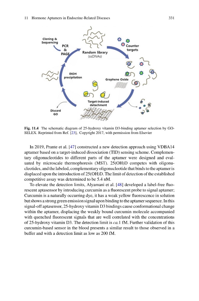
Fig. 11.4 The schematic diagram of 25-hydroxy vitamin D3-binding aptamer selection by GOSELEX. Reprinted from Ref. [23], Copyright 2017, with permission from Elsevier
In 2019, Prante et al. [47] constructed a new detection approach using VDBA14
aptamer based on a target-induced dissociation (TID) sensing scheme. Complementary oligonucleotides to different parts of the aptamer were designed and evaluated by microscale thermophoresis (MST). 25(OH)D competes with oligonucleotides, and the labeled, complementary oligonucleotide that binds to the aptamer is
displaced upon the introduction of 25(OH)D. The limit of detection of the established
competitive assay was determined to be 5.4 nM.
To elevate the detection limits, Alyamani et al. [48] developed a label-free fluorescent aptasensor by introducing curcumin as a fluorescent probe to signal aptamer;
Curcumin is a naturally occurring dye, it has a weak yellow fluorescence in solution
but shows a strong green emission signal upon binding to the aptamer sequence. In this
signal-off aptasensor, 25-hydroxy vitamin D3 bindings cause conformational change
within the aptamer, displacing the weakly bound curcumin molecule accompanied
with quenched fluorescent signals that are well correlated with the concentrations
of 25-hydroxy vitamin D3. The detection limit is ca.1 fM. Further validation of this
curcumin-based sensor in the blood presents a similar result to those observed in a
buffer and with a detection limit as low as 200 fM.
331
Fig. 11.4 The schematic diagram of 25-hydroxy vitamin D3-binding aptamer selection by GOSELEX. Reprinted from Ref. [23], Copyright 2017, with permission from Elsevier
In 2019, Prante et al. [47] constructed a new detection approach using VDBA14
aptamer based on a target-induced dissociation (TID) sensing scheme. Complementary oligonucleotides to different parts of the aptamer were designed and evaluated by microscale thermophoresis (MST). 25(OH)D competes with oligonucleotides, and the labeled, complementary oligonucleotide that binds to the aptamer is
displaced upon the introduction of 25(OH)D. The limit of detection of the established
competitive assay was determined to be 5.4 nM.
To elevate the detection limits, Alyamani et al. [48] developed a label-free fluorescent aptasensor by introducing curcumin as a fluorescent probe to signal aptamer;
Curcumin is a naturally occurring dye, it has a weak yellow fluorescence in solution
but shows a strong green emission signal upon binding to the aptamer sequence. In this
signal-off aptasensor, 25-hydroxy vitamin D3 bindings cause conformational change
within the aptamer, displacing the weakly bound curcumin molecule accompanied
with quenched fluorescent signals that are well correlated with the concentrations
of 25-hydroxy vitamin D3. The detection limit is ca.1 fM. Further validation of this
curcumin-based sensor in the blood presents a similar result to those observed in a
buffer and with a detection limit as low as 200 fM.