

328
Y. Cheng and H. Jin
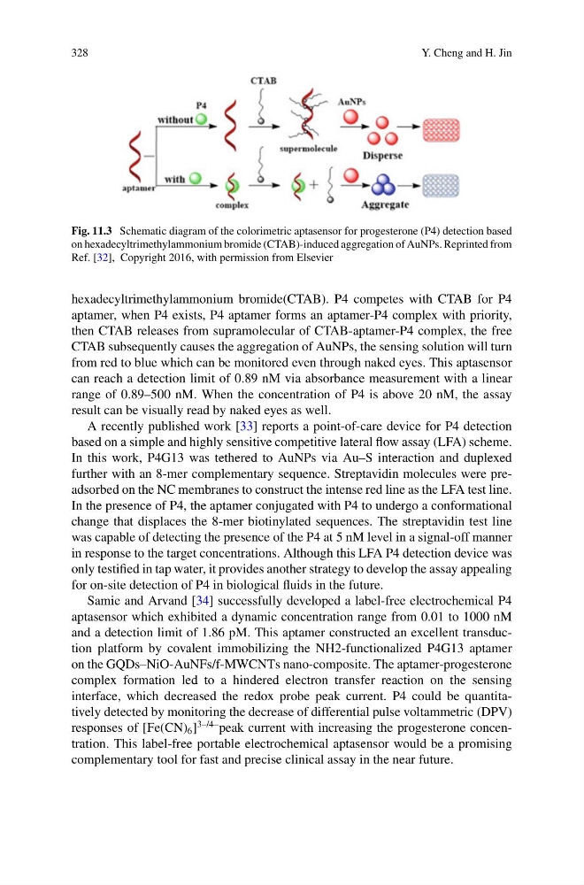
Fig. 11.3 Schematic diagram of the colorimetric aptasensor for progesterone (P4) detection based
on hexadecyltrimethylammonium bromide (CTAB)-induced aggregation of AuNPs. Reprinted from
Ref. [32], Copyright 2016, with permission from Elsevier
hexadecyltrimethylammonium bromide(CTAB). P4 competes with CTAB for P4
aptamer, when P4 exists, P4 aptamer forms an aptamer-P4 complex with priority,
then CTAB releases from supramolecular of CTAB-aptamer-P4 complex, the free
CTAB subsequently causes the aggregation of AuNPs, the sensing solution will turn
from red to blue which can be monitored even through naked eyes. This aptasensor
can reach a detection limit of 0.89 nM via absorbance measurement with a linear
range of 0.89–500 nM. When the concentration of P4 is above 20 nM, the assay
result can be visually read by naked eyes as well.
A recently published work [33] reports a point-of-care device for P4 detection
based on a simple and highly sensitive competitive lateral flow assay (LFA) scheme.
In this work, P4G13 was tethered to AuNPs via Au–S interaction and duplexed
further with an 8-mer complementary sequence. Streptavidin molecules were preadsorbed on the NC membranes to construct the intense red line as the LFA test line.
In the presence of P4, the aptamer conjugated with P4 to undergo a conformational
change that displaces the 8-mer biotinylated sequences. The streptavidin test line
was capable of detecting the presence of the P4 at 5 nM level in a signal-off manner
in response to the target concentrations. Although this LFA P4 detection device was
only testified in tap water, it provides another strategy to develop the assay appealing
for on-site detection of P4 in biological fluids in the future.
Samie and Arvand [34] successfully developed a label-free electrochemical P4
aptasensor which exhibited a dynamic concentration range from 0.01 to 1000 nM
and a detection limit of 1.86 pM. This aptamer constructed an excellent transduction platform by covalent immobilizing the NH2-functionalized P4G13 aptamer
on the GQDs–NiO-AuNFs/f-MWCNTs nano-composite. The aptamer-progesterone
complex formation led to a hindered electron transfer reaction on the sensing
interface, which decreased the redox probe peak current. P4 could be quantitatively detected by monitoring the decrease of differential pulse voltammetric (DPV)
responses of [Fe(CN) 6 ]
3–/4– peak current with increasing the progesterone concentration. This label-free portable electrochemical aptasensor would be a promising
complementary tool for fast and precise clinical assay in the near future.
Y. Cheng and H. Jin
Fig. 11.3 Schematic diagram of the colorimetric aptasensor for progesterone (P4) detection based
on hexadecyltrimethylammonium bromide (CTAB)-induced aggregation of AuNPs. Reprinted from
Ref. [32], Copyright 2016, with permission from Elsevier
hexadecyltrimethylammonium bromide(CTAB). P4 competes with CTAB for P4
aptamer, when P4 exists, P4 aptamer forms an aptamer-P4 complex with priority,
then CTAB releases from supramolecular of CTAB-aptamer-P4 complex, the free
CTAB subsequently causes the aggregation of AuNPs, the sensing solution will turn
from red to blue which can be monitored even through naked eyes. This aptasensor
can reach a detection limit of 0.89 nM via absorbance measurement with a linear
range of 0.89–500 nM. When the concentration of P4 is above 20 nM, the assay
result can be visually read by naked eyes as well.
A recently published work [33] reports a point-of-care device for P4 detection
based on a simple and highly sensitive competitive lateral flow assay (LFA) scheme.
In this work, P4G13 was tethered to AuNPs via Au–S interaction and duplexed
further with an 8-mer complementary sequence. Streptavidin molecules were preadsorbed on the NC membranes to construct the intense red line as the LFA test line.
In the presence of P4, the aptamer conjugated with P4 to undergo a conformational
change that displaces the 8-mer biotinylated sequences. The streptavidin test line
was capable of detecting the presence of the P4 at 5 nM level in a signal-off manner
in response to the target concentrations. Although this LFA P4 detection device was
only testified in tap water, it provides another strategy to develop the assay appealing
for on-site detection of P4 in biological fluids in the future.
Samie and Arvand [34] successfully developed a label-free electrochemical P4
aptasensor which exhibited a dynamic concentration range from 0.01 to 1000 nM
and a detection limit of 1.86 pM. This aptamer constructed an excellent transduction platform by covalent immobilizing the NH2-functionalized P4G13 aptamer
on the GQDs–NiO-AuNFs/f-MWCNTs nano-composite. The aptamer-progesterone
complex formation led to a hindered electron transfer reaction on the sensing
interface, which decreased the redox probe peak current. P4 could be quantitatively detected by monitoring the decrease of differential pulse voltammetric (DPV)
responses of [Fe(CN) 6 ]
3–/4– peak current with increasing the progesterone concentration. This label-free portable electrochemical aptasensor would be a promising
complementary tool for fast and precise clinical assay in the near future.