

266
Q. Lv et al.
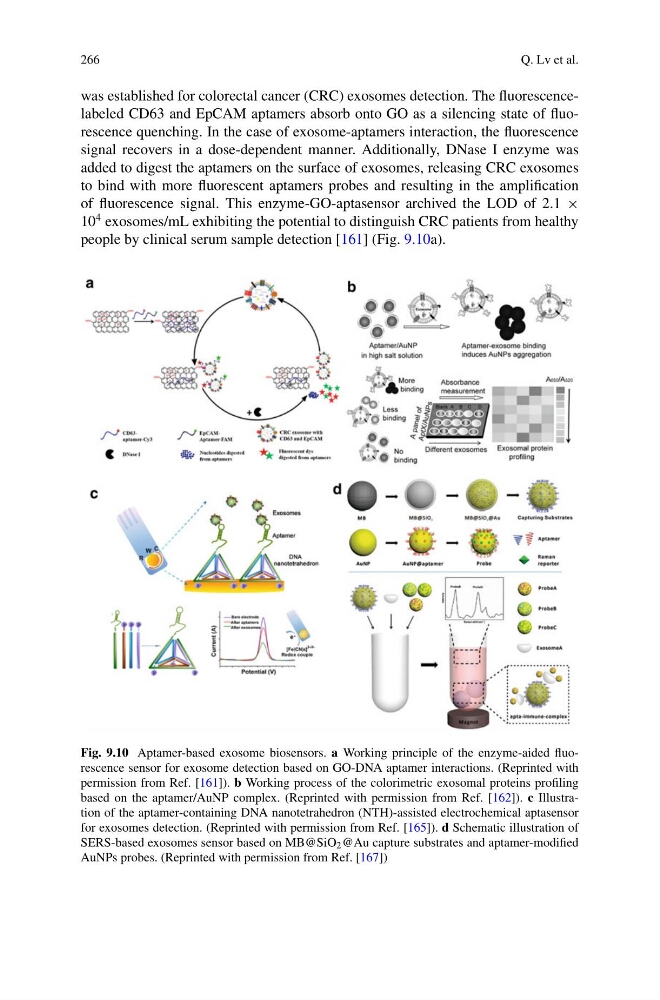
was established for colorectal cancer (CRC) exosomes detection. The fluorescencelabeled CD63 and EpCAM aptamers absorb onto GO as a silencing state of fluorescence quenching. In the case of exosome-aptamers interaction, the fluorescence
signal recovers in a dose-dependent manner. Additionally, DNase I enzyme was
added to digest the aptamers on the surface of exosomes, releasing CRC exosomes
to bind with more fluorescent aptamers probes and resulting in the amplification
of fluorescence signal. This enzyme-GO-aptasensor archived the LOD of 2.1 ×
10
4 exosomes/mL exhibiting the potential to distinguish CRC patients from healthy
people by clinical serum sample detection [161] (Fig. 9.10a).
Fig. 9.10 Aptamer-based exosome biosensors. a Working principle of the enzyme-aided fluorescence sensor for exosome detection based on GO-DNA aptamer interactions. (Reprinted with
permission from Ref. [161]). b Working process of the colorimetric exosomal proteins profiling
based on the aptamer/AuNP complex. (Reprinted with permission from Ref. [162]). c Illustration of the aptamer-containing DNA nanotetrahedron (NTH)-assisted electrochemical aptasensor
for exosomes detection. (Reprinted with permission from Ref. [165]). d Schematic illustration of
SERS-based exosomes sensor based on MB@SiO 2 @Au capture substrates and aptamer-modified
AuNPs probes. (Reprinted with permission from Ref. [167])
Q. Lv et al.
was established for colorectal cancer (CRC) exosomes detection. The fluorescencelabeled CD63 and EpCAM aptamers absorb onto GO as a silencing state of fluorescence quenching. In the case of exosome-aptamers interaction, the fluorescence
signal recovers in a dose-dependent manner. Additionally, DNase I enzyme was
added to digest the aptamers on the surface of exosomes, releasing CRC exosomes
to bind with more fluorescent aptamers probes and resulting in the amplification
of fluorescence signal. This enzyme-GO-aptasensor archived the LOD of 2.1 ×
10
4 exosomes/mL exhibiting the potential to distinguish CRC patients from healthy
people by clinical serum sample detection [161] (Fig. 9.10a).
Fig. 9.10 Aptamer-based exosome biosensors. a Working principle of the enzyme-aided fluorescence sensor for exosome detection based on GO-DNA aptamer interactions. (Reprinted with
permission from Ref. [161]). b Working process of the colorimetric exosomal proteins profiling
based on the aptamer/AuNP complex. (Reprinted with permission from Ref. [162]). c Illustration of the aptamer-containing DNA nanotetrahedron (NTH)-assisted electrochemical aptasensor
for exosomes detection. (Reprinted with permission from Ref. [165]). d Schematic illustration of
SERS-based exosomes sensor based on MB@SiO 2 @Au capture substrates and aptamer-modified
AuNPs probes. (Reprinted with permission from Ref. [167])