

260
Q. Lv et al.
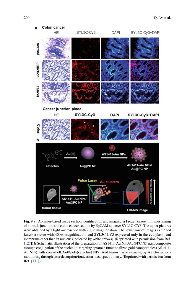
Fig. 9.8 Aptamer-based tissue section identification and imaging. a Frozen tissue immunostaining
of normal, junction, and colon cancer section by EpCAM aptamer SYL3C-CY3. The upper pictures
were obtained by a light microscope with 200× magnification. The lower row of images exhibited
junction tissue with 400× magnification, and SYL3C-CY3 expressed only in the cytoplasm and
membrane other than in nucleus (indicated by white arrows). (Reprinted with permission from Ref.
[127]) b Schematic illustration of the preparation of AS1411-Au NPs/Au@PC NP nanocomposite
through conjugation of the nucleolin-targeting aptamer-functionalized gold nanoparticles (AS1411Au NPs) with core-shell Au@poly(catechin) NPs. And tumor tissue imaging by Au cluster ions
monitoring through laser desorption/ionization mass spectrometry. (Reprinted with permission from
Ref. [131])
Q. Lv et al.
Fig. 9.8 Aptamer-based tissue section identification and imaging. a Frozen tissue immunostaining
of normal, junction, and colon cancer section by EpCAM aptamer SYL3C-CY3. The upper pictures
were obtained by a light microscope with 200× magnification. The lower row of images exhibited
junction tissue with 400× magnification, and SYL3C-CY3 expressed only in the cytoplasm and
membrane other than in nucleus (indicated by white arrows). (Reprinted with permission from Ref.
[127]) b Schematic illustration of the preparation of AS1411-Au NPs/Au@PC NP nanocomposite
through conjugation of the nucleolin-targeting aptamer-functionalized gold nanoparticles (AS1411Au NPs) with core-shell Au@poly(catechin) NPs. And tumor tissue imaging by Au cluster ions
monitoring through laser desorption/ionization mass spectrometry. (Reprinted with permission from
Ref. [131])