

250
Q. Lv et al.
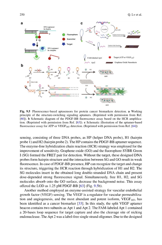
Fig. 9.5 Fluorescence-based aptasensors for protein cancer biomarkers detection. a Working
principle of the structure-switching signaling aptamers. (Reprinted with permission from Ref.
[60]). b Schematic diagram of the PDGF-BB fluorescence assay based on the HCR amplification. (Reprinted with permission from Ref. [63]). c Schematic illustration of the aptamer-based
fluorescence assay for ATP or VEGF 165 detection. (Reprinted with permission from Ref. [64])
sensing, consisting of three DNA probes, as HP (helper DNA probe), H1 (hairpin
probe 1) and H2 (hairpin probe 2). The HP contains the PDGF-BB aptamer sequence.
The enzyme-free hybridization chain reaction (HCR) strategy was employed for the
improvement of sensitivity. Graphene oxide (GO) and the fluorophore SYBR Green
I (SG) formed the FRET pair for detection. Without the target, these designed DNA
probes form hairpin structure and the interaction between SG and GO result in weak
fluorescence. In case of PDGF-BB presence, HP can recognize the target and change
its structure, triggering the HCR reaction through hybridization of H1 and H2. The
SG molecules insert in the obtained long double-stranded DNA chain and present
dose-depended strong fluorescence signal. Simultaneously, free H1, H2, and SG
molecules absorb onto the GO surface, decrease the background noise. This assay
offered the LOD as 1.25 pM PDGF-BB [63] (Fig. 9.5b).
Another method employed an enzyme-assisted strategy for vascular endothelial
growth factor (VEGF) sensing. The VEGF is a regulator for vascular permeabilization and angiogenesis, and the most abundant and potent isoform, VEGF 165 , has
been identified as a cancer biomarker [33]. In this study, the split VEGF aptamer
beacon contains two subunits as Apt 1 and Apt 2. The FAM-labeled Apt 1 contained
a 20-bases loop sequence for target capture and also the cleavage site of nicking
endonuclease. The Apt 2 was a label-free single strand oligomer. Due to the designed
Q. Lv et al.
Fig. 9.5 Fluorescence-based aptasensors for protein cancer biomarkers detection. a Working
principle of the structure-switching signaling aptamers. (Reprinted with permission from Ref.
[60]). b Schematic diagram of the PDGF-BB fluorescence assay based on the HCR amplification. (Reprinted with permission from Ref. [63]). c Schematic illustration of the aptamer-based
fluorescence assay for ATP or VEGF 165 detection. (Reprinted with permission from Ref. [64])
sensing, consisting of three DNA probes, as HP (helper DNA probe), H1 (hairpin
probe 1) and H2 (hairpin probe 2). The HP contains the PDGF-BB aptamer sequence.
The enzyme-free hybridization chain reaction (HCR) strategy was employed for the
improvement of sensitivity. Graphene oxide (GO) and the fluorophore SYBR Green
I (SG) formed the FRET pair for detection. Without the target, these designed DNA
probes form hairpin structure and the interaction between SG and GO result in weak
fluorescence. In case of PDGF-BB presence, HP can recognize the target and change
its structure, triggering the HCR reaction through hybridization of H1 and H2. The
SG molecules insert in the obtained long double-stranded DNA chain and present
dose-depended strong fluorescence signal. Simultaneously, free H1, H2, and SG
molecules absorb onto the GO surface, decrease the background noise. This assay
offered the LOD as 1.25 pM PDGF-BB [63] (Fig. 9.5b).
Another method employed an enzyme-assisted strategy for vascular endothelial
growth factor (VEGF) sensing. The VEGF is a regulator for vascular permeabilization and angiogenesis, and the most abundant and potent isoform, VEGF 165 , has
been identified as a cancer biomarker [33]. In this study, the split VEGF aptamer
beacon contains two subunits as Apt 1 and Apt 2. The FAM-labeled Apt 1 contained
a 20-bases loop sequence for target capture and also the cleavage site of nicking
endonuclease. The Apt 2 was a label-free single strand oligomer. Due to the designed