

248
Q. Lv et al.
problem, several nanoparticles are utilized in SPR biosensors construction for further
signal amplification. Nanoparticle-assisted SPR biosensors have been employed for
detecting various cancer biomarkers, such as carcinoembryonic antigen (CEA) [46],
human epidermal growth factor receptor 2 (HER2) [47, 48], total prostate-specific
antigen (tPSA) [49], platelet-derived growth factor (PDGF-BB) [50], progesterone
receptor (PR) [51], carbohydrate antigen 15-3 (CA15-3) [52], etc. CEA is a common
serologic biomarker for the diagnosis of gastric, colorectal, and pancreatic cancers.
The CEA content in serum samples of a healthy person is below 5 ng/mL. An
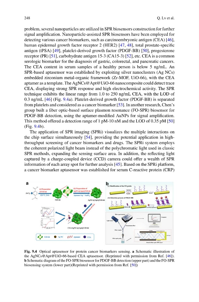
SPR-based aptasensor was established by exploiting silver nanoclusters (Ag NCs)
embedded zirconium metal-organic framework (Zr-MOF, UiO-66), with the CEA
aptamer as a template. The AgNCs@Apt@UiO-66 nanocomposite could detect trace
CEA, displaying strong SPR response and high electrochemical activity. The SPR
technique exhibits the linear range from 1.0 to 250 ng/mL CEA, with the LOD of
0.3 ng/mL [46] (Fig. 9.4a). Platelet-derived growth factor (PDGF-BB) is separated
from platelets and considered as a cancer biomarker [53]. In another research, Chen’s
group built a fiber optic-based surface plasmon resonance (FO-SPR) biosensor for
PDGF-BB detection, using the aptamer-modified AuNPs for signal amplification.
This method offered a detection range of 1 pM-10 nM and the LOD of 0.35 pM [50]
(Fig. 9.4b).
The application of SPR imaging (SPRi) visualizes the multiple interactions on
the chip surface simultaneously [54], providing the potential application in highthroughput screening of cancer biomarkers and drugs. The SPRi system employs
the coherent polarized light beam instead of the polychromatic light used in classic
SPR methods, expanding the sensing surface area. In addition, the reflecting light
captured by a charge-coupled device (CCD) camera could offer a wealth of SPR
information of each array spot for further analysis [45]. Based on the SPRi platform,
a cancer biomarker aptasensor was established for serum C-reactive protein (CRP)
Fig. 9.4 Optical aptasensor for protein cancer biomarkers sensing. a Schematic illustration of
the AgNCs@Apt@UiO-66-based CEA aptasensor. (Reprinted with permission from Ref. [46]).
b Schematic diagram of the FO-SPR biosensor for PDGF-BB detection (upper part) and the FO-SPR
biosensing system (lower part)(Reprinted with permission from Ref. [50])
Q. Lv et al.
problem, several nanoparticles are utilized in SPR biosensors construction for further
signal amplification. Nanoparticle-assisted SPR biosensors have been employed for
detecting various cancer biomarkers, such as carcinoembryonic antigen (CEA) [46],
human epidermal growth factor receptor 2 (HER2) [47, 48], total prostate-specific
antigen (tPSA) [49], platelet-derived growth factor (PDGF-BB) [50], progesterone
receptor (PR) [51], carbohydrate antigen 15-3 (CA15-3) [52], etc. CEA is a common
serologic biomarker for the diagnosis of gastric, colorectal, and pancreatic cancers.
The CEA content in serum samples of a healthy person is below 5 ng/mL. An
SPR-based aptasensor was established by exploiting silver nanoclusters (Ag NCs)
embedded zirconium metal-organic framework (Zr-MOF, UiO-66), with the CEA
aptamer as a template. The AgNCs@Apt@UiO-66 nanocomposite could detect trace
CEA, displaying strong SPR response and high electrochemical activity. The SPR
technique exhibits the linear range from 1.0 to 250 ng/mL CEA, with the LOD of
0.3 ng/mL [46] (Fig. 9.4a). Platelet-derived growth factor (PDGF-BB) is separated
from platelets and considered as a cancer biomarker [53]. In another research, Chen’s
group built a fiber optic-based surface plasmon resonance (FO-SPR) biosensor for
PDGF-BB detection, using the aptamer-modified AuNPs for signal amplification.
This method offered a detection range of 1 pM-10 nM and the LOD of 0.35 pM [50]
(Fig. 9.4b).
The application of SPR imaging (SPRi) visualizes the multiple interactions on
the chip surface simultaneously [54], providing the potential application in highthroughput screening of cancer biomarkers and drugs. The SPRi system employs
the coherent polarized light beam instead of the polychromatic light used in classic
SPR methods, expanding the sensing surface area. In addition, the reflecting light
captured by a charge-coupled device (CCD) camera could offer a wealth of SPR
information of each array spot for further analysis [45]. Based on the SPRi platform,
a cancer biomarker aptasensor was established for serum C-reactive protein (CRP)
Fig. 9.4 Optical aptasensor for protein cancer biomarkers sensing. a Schematic illustration of
the AgNCs@Apt@UiO-66-based CEA aptasensor. (Reprinted with permission from Ref. [46]).
b Schematic diagram of the FO-SPR biosensor for PDGF-BB detection (upper part) and the FO-SPR
biosensing system (lower part)(Reprinted with permission from Ref. [50])