

226
T. Zhang et al.
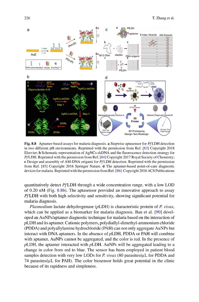
Fig. 8.8 Aptamer-based assays for malaria diagnosis. a Stepwise aptasensor for Pf LDH detection
in two different pH environments. Reprinted with the permission from Ref. [83] Copyright 2018
Elsevier; b Schematic representation of AgNCs-dsDNA and the fluorescence detection strategy for
Pf LDH. Reprinted with the permission from Ref. [84] Copyright 2017 Royal Society of Chemistry;
c Design and assembly of AM-DNA origami for Pf LDH detection. Reprinted with the permission
from Ref. [85] Copyright 2016 Springer Nature. d The aptamer-based point-of-care diagnostic
devices for malaria. Reprinted with the permission from Ref. [86]. Copyright 2016 ACS Publications
quantitatively detect Pf LDH through a wide concentration range, with a low LOD
of 0.20 nM (Fig. 8.8b). The aptasensor provided an innovative approach to assay
Pf LDH with both high selectivity and sensitivity, showing significant potential for
malaria diagnosis.
Plasmodium lactate dehydrogenase (pLDH) is characteristic protein of P. vivax,
which can be applied as a biomarker for malaria diagnosis. Ban et al. [90] developed an AuNPs/aptamer diagnostic technique for malaria based on the interaction of
pLDH and its aptamer. Cationic polymers, polydiallyl-dimethyl-ammonium chloride
(PDDA) and polyallylamine hydrochloride (PAH) can not only aggregate AuNPs but
interact with DNA aptamers. In the absence of pLDH, PDDA or PAH will combine
with aptamer, AuNPs cannot be aggregated, and the color is red. In the presence of
pLDH, the aptamer interacted with pLDH. AuNPs will be aggregated leading to a
change in color from red to blue. The sensor has been employed in patient blood
samples detection with very low LODs for P. vivax (80 parasites/µL for PDDA and
74 parasites/µL for PAH). The color biosensor holds great potential in the clinic
because of its rapidness and simpleness.
T. Zhang et al.
Fig. 8.8 Aptamer-based assays for malaria diagnosis. a Stepwise aptasensor for Pf LDH detection
in two different pH environments. Reprinted with the permission from Ref. [83] Copyright 2018
Elsevier; b Schematic representation of AgNCs-dsDNA and the fluorescence detection strategy for
Pf LDH. Reprinted with the permission from Ref. [84] Copyright 2017 Royal Society of Chemistry;
c Design and assembly of AM-DNA origami for Pf LDH detection. Reprinted with the permission
from Ref. [85] Copyright 2016 Springer Nature. d The aptamer-based point-of-care diagnostic
devices for malaria. Reprinted with the permission from Ref. [86]. Copyright 2016 ACS Publications
quantitatively detect Pf LDH through a wide concentration range, with a low LOD
of 0.20 nM (Fig. 8.8b). The aptasensor provided an innovative approach to assay
Pf LDH with both high selectivity and sensitivity, showing significant potential for
malaria diagnosis.
Plasmodium lactate dehydrogenase (pLDH) is characteristic protein of P. vivax,
which can be applied as a biomarker for malaria diagnosis. Ban et al. [90] developed an AuNPs/aptamer diagnostic technique for malaria based on the interaction of
pLDH and its aptamer. Cationic polymers, polydiallyl-dimethyl-ammonium chloride
(PDDA) and polyallylamine hydrochloride (PAH) can not only aggregate AuNPs but
interact with DNA aptamers. In the absence of pLDH, PDDA or PAH will combine
with aptamer, AuNPs cannot be aggregated, and the color is red. In the presence of
pLDH, the aptamer interacted with pLDH. AuNPs will be aggregated leading to a
change in color from red to blue. The sensor has been employed in patient blood
samples detection with very low LODs for P. vivax (80 parasites/µL for PDDA and
74 parasites/µL for PAH). The color biosensor holds great potential in the clinic
because of its rapidness and simpleness.